. 2009 Sep 15;162(4):989-1000.
doi: 10.1016/j.neuroscience.2009.05.016. Epub 2009 May 14.

Large conductance calcium-activated potassium channels affect both spontaneous firing and intracellular calcium concentration in cerebellar Purkinje neurons

Affiliations

Large conductance calcium-activated potassium channels affect both spontaneous firing and intracellular calcium concentration in cerebellar Purkinje neurons

M D Womack et al. Neuroscience. .

Abstract

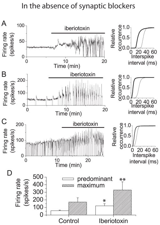
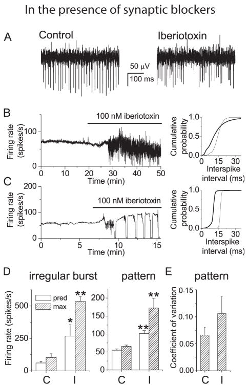
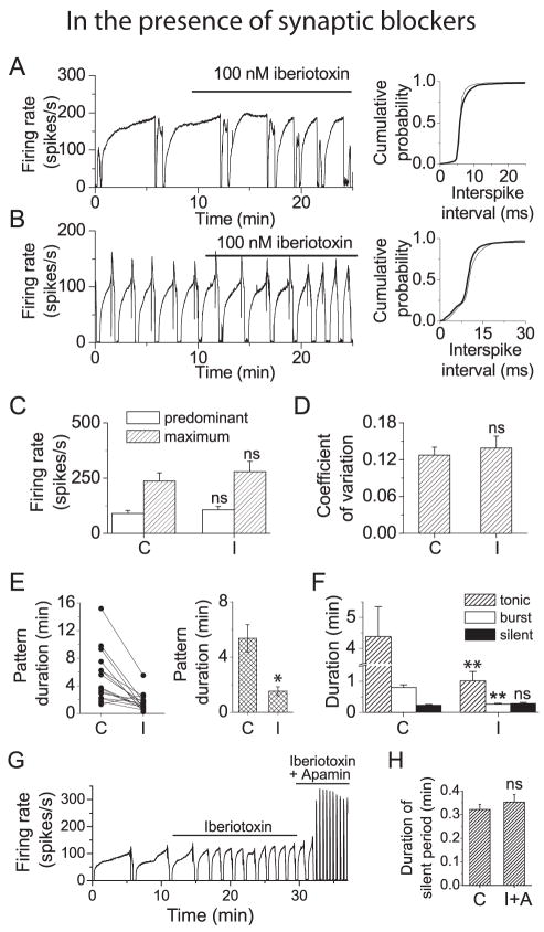
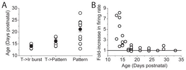
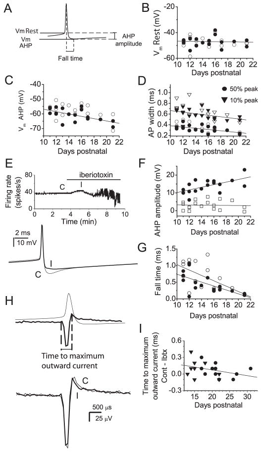
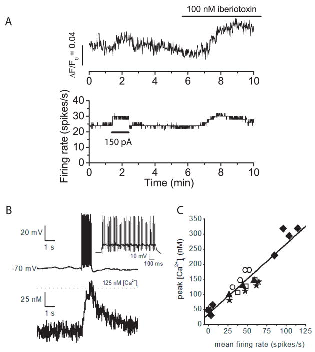
We investigated the contribution of large conductance calcium-activated potassium (BK) channels to spontaneous activity of cerebellar Purkinje neurons in mice and rats. In Purkinje neurons which fire tonically, block of BK channels increased the firing rate and caused the neurons to fire irregularly. In Purkinje neurons which exhibited a trimodal pattern of activity, present primarily in mature animals, block of BK channels had little effect on firing rate or regularity but shortened the single cycle duration of the trimodal pattern. The contribution of BK channels to the action potential waveform was also examined. BK channels contributed a brief afterhyperpolarization (AHP) of approximately 3 mV which followed each action potential, but made little contribution to action potential repolarization. The amplitude of the BK-dependent AHP did not change with age although there was an increase in the total AHP. The difference in the contribution of BK channels to the firing rate among the two populations of Purkinje neurons was the consequence of the decrease in the fractional contribution of BK channels to the AHP. We also found that block of BK channels increases intracellular calcium concentration during spontaneous firing. Thus, although BK channels do not affect action potential repolarization, they nevertheless control calcium entry with each action potential by contributing to the AHP.


Figures

Figure 1
Figure 1. Block of BK channels increases firing rate and promotes bursting in cerebellar Purkinje neurons
A, Left panel: Average firing rate (calculated every 500 ms) vs. time recorded from a Purkinje neuron from a 14 day old rat which fired tonically. Superfusion with iberiotoxin (100 nM) increased the firing rate and caused the cell to fire irregularly. The cumulative distribution of interspike intervals shows a shift to shorter intervals (higher firing rate) in iberiotoxin (right panel). Thin trace, control distribution; thick trace, distribution in iberiotoxin. B, Left panel: Average firing rate vs. time from a Purkinje neuron from a 21 day old rat which exhibited the trimodal pattern of activity. Application of iberiotoxin increased the firing rate and increased the fraction of time that the cell was in the bursting mode. Right panel: Cumulative probability distribution s of interspike intervals under control conditions (thin traces) and in iberiotoxin (thick traces). C, Left panel: Average firing rate vs. time from a Purkinje neuron from a 18 day old rat which burst at random. Application of iberiotoxin increased the firing rate. Right panel: Cumulative probability distribution s of interspike intervals under control conditions (thin traces) and in iberiotoxin (thick traces). D, Mean predominant and maximum firing rates under control conditions and in the presence of iberiotoxin. Predominant and maximum firing rates for each cell were determined from histograms of interspike intervals. The predominant firing rate was defined as the firing rate most often observed (peak of the histogram). The maximum firing rate was the fastest firing rate observed at 5% of the frequency of the predominant rate. Error bars are ± SEM (n=5). Under control conditions the mean predominant and maximum firing rates were 58 ± 8 and 171 ± 56 spikes per second, respectively. In the presence of iberiotoxin the mean predominant firing rate increased to 121 ± 29 spikes per second while the maximum firing rate was 330 ±108 spikes per second. Error bars are mean ± S.E.M. (n=5). Control values were significantly different from those in iberiotoxin (*, p< = 0.07; ** p< = 0.05 by Oneway ANOVA).
Figure 2
Figure 2. In tonic firing neurons block of BK channels increases firing rate and promotes bursting
A, Extracellular recordings from a Purkinje neuron which fired tonically under control conditions in the presence of synaptic blockers (left trace). Superfusion with iberiotoxin (100 nM) caused the cell to fire irregularly (right trace). B, Left panel: Average firing rate vs. time from the neuron discussed in A. Right panel: Cumulative probability distributions of interspike intervals in the presence of iberiotoxin (thick traces) and in control conditions (thin traces) show that iberiotoxin shifted the distribution to shorter intervals (higher firing rate). C, Left panel: Average firing rate vs. time from another tonically firing Purkinje neuron. Superfusion with iberiotoxin increased the firing rate and induced the trimodal pattern of activity. Right panel: Cumulative probability distributions of interspike intervals in the presence of iberiotoxin (thick traces) and in control conditions (thin traces). D, Mean predominant and maximum firing rates for cells in which iberiotoxin caused irregular bursting (left panel) and for cells in which iberiotoxin caused the trimodal pattern of activity (middle panel). In irregular bursting cells block of BK channels significantly increased the predominant firing rate from 61 (± 10) spikes/s in control conditions (C) to 268 (± 87) in iberiotoxin (I) and the maximum firing rate from 102 (± 29) spikes/s to 536 (± 36) spikes/s. In Neurons which acquired the trimodal pattern, block of BK channels significantly increased the predominant firing rate from 55 (± 3) spikes/s to 102 (± 29) and the maximum firing rate from 66 (± 4) spikes/s to 173 (± 28) spikes/s. Values in iberiotoxin were significantly different from control for (*) p<=0.05 and (**) p<= 0.005 (One-way ANOVA). Error bars are mean ± S.E.M. (n=5 for each type of neuron). E, For cells in which iberiotoxin induced a trimodal pattern of firing, the coefficient of variation (CV) for interspike intervals was measured under control conditions and in the presence of iberiotoxin during a period of the tonic phase of the trimodal pattern. Block of BK channels increased the CV from 0.066 (± 0.01) to 0.11 (± 0.03). Error bars are ± S.E.M. (n=5).
Figure 3
Figure 3. In Purkinje neurons with the trimodal pattern of activity block of BK channels shortens the pattern period but does not increase the average firing rate
A, Left panel: Average firing rate vs. time from a Purkinje neuron with the trimodal pattern of activity. Superfusion with iberiotoxin decreased the single cycle duration of the pattern period but did not increase the average firing rate. Right panel: Cumulative probability distributions of interspike intervals in the presence of iberiotoxin (thick traces) and in control conditions (thin traces) show that there was no change in firing rate in iberiotoxin. B, Left panel: Average firing rate vs. time from another Purkinje neuron with the trimodal pattern. Right panel: Cumulative probability distributions of interspike intervals. C, Histogram showing average predominant and maximum firing rates for all Purkinje neurons with the trimodal pattern. The mean value of the predominant firing rate was 90 (± 14) spikes/s in control conditions (C) and 107 (± 15) spikes/s in iberiotoxin (I). Maximum firing rates were 237 (± 37) spikes/s in control conditions, and 279 (± 47) spikes/s in iberiotoxin, respectively. Error bars are ± S.E.M., n=15. ns, values were not significantly different from control. D, The coefficient of variation was determined for a 5 s interval where there was no increase in the average firing rate during the tonic phase of firing for each neuron. Block of BK channels did not change the coefficient of variation. mean values were o.13 (± 0.01) in control conditions and 0.14 (± 0.02) in iberiotoxin. ns, values were not significantly different from control. E, Left panel: The average single cycle duration for each cell tested under control conditions (C) and in the presence of iberiotoxin (I). Right panel: Average single cycle duration for all cells tested. The mean pattern duration was 5.36 (± 1.0) min under control conditions and 1.53 (± 0.3) min in iberiotoxin. Error bars are ± S.E.M (n = 15). Values in iberiotoxin were significantly different from control (*, p = 0.001). F, Histogram showing the average duration of the tonic, bursting, and silent periods in control conditions and in iberiotoxin. Block of BK channels decreased the average duration of the tonic firing mode from 4.37 (± 1.0) min to 1.0 (± 0.3) min and the duration of the bursting mode from 0.79 (± 0.09) to 0.26 (± 0.03) min. (**, differences were significant at p<=0.005). There was no significant change in the duration of the silent period (0.23 (± 0.03) min in control vs. 0.28 (± 0.03) min in iberiotoxin (ns). Error bars are ± S.E.M. (n = 15) G, Average firing rate vs. time following application of iberiotoxin (100 nM) and simultaneous application of iberiotoxin and the specific SK channel blocker apamin (100 nM) to a neurons with the trimodal pattern. In the presence of both blockers the neuron continues to fire with the trimodal pattern. H, Histogram comparing the average duration of the silent period in control conditions (C) and in the presence of iberiotoxin and apamin (I+A). Simultaneous block of both BK and SK channels did not alter the duration of the silent period (0.32 (±0.02) min in control vs. 0.35 (±0.03) min. in iberiotoxin + apamin). ns, values not significantly different from control. Error bars are ± S.E.M. (n = 5).
Figure 4
Figure 4. BK channels which control firing rate are localized to the soma of Purkinje neurons while BK channels which regulate the duration of the trimodal pattern are localized to both soma and dendrites
Iberiotoxin (100 nM) was applied selectively via local perfusion to the distal half (1/2 D) or distal two thirds (2/3 D) of the dendrites or to the whole cell (WC). A, Average firing rate of a tonic firing neuron in response to local application of iberiotoxin. Application to the dendrites had no effect. B, Average firing rate of a neuron with the trimodal pattern of activity in response to local application of iberiotoxin. C, Average fold-increase in firing rate of tonic firing neurons following local block of BK channels. Block of CK channels to the distal ½ (no effect) or distal 2/3 of the dendrites (0.11 (± 0.02)-fold increase) did not significantly change the firing rate. Block of BK channels I the whole cell increased the firing rate by and average of 1.28 (± 0.14)-fold. Error bars are ± S.E.M., n=4. ns, value not significantly different from control. **, significantly different from 0 (p< = 0.001, by Oneway ANOVA). D, Average fold-increase in firing rate (hatched bars) and fold- decrease in pattern duration (open bars) in Purkinje neurons with the trimodal pattern of activity. Block of BK channels did not significantly increase firing rate. The mean fold-increases in firing rate were (1/2 D,) −0.05 (± 0.07), (2/3 D), 0.09 (± 0.03), and (WC), 0.04 (± 0.09). None of these values was significantly different from 0 (ns). Block of BK channels in the dendrites decreased the pattern period. Mean fold- decreases were 0.24 (± 0.08) (1/2 D), and 0.5 (± 0.03) (2/3 D). Application to the whole cell decreased the period 0.84 (± 0.04)-fold. Error bars are ± S.E.M., n=7. The changes were significant (*, p = 0.02, **, p = 0.001 by Oneway ANOVA).
Figure 5
Figure 5. Age-dependence of effects of Iberiotoxin on spontaneoeuos firing of Purkinje cells
A,Open symbols show the age of animals in which iberiotoxin altered tonic firing to irregular burst firing (T->Ir Burst; n=7), tonic firing to the trimodal pattern of activity (T->Pattern; n=6), and cells that in the absence of iberiotoxin exhibited the trimodal modal pattern of activity (Pattern; n=15). Filled symbols show the average age (±S.E.M.). The average ages of the three groups were significantly different from each other. B, The relationship between the age of animals and the extent to which iberiotoxin increased their firing rate.
Figure 6
Figure 6. Contribution of BK channels to action potential waveform
A–G, Whole cell current clamp recordings were obtained from spontaneously firing Purkinje neurons. Trains of 100–1500 action potentials were averaged and action potential waveforms analyzed from the average traces. A, The diagram shows parameters measured from the average action potentials. Vm Rest, membrane potential 500 μs prior to the peak of the action potential; Vm AHP, minimum membrane potential following action potential; AHP amplitude, Vm Rest - Vm AHP; fall time, time from peak of action potential to Vm AHP. The action potential width at 10 %and 50 % of the difference between the peak and Vm Rest was also measured. B, Vm Rest vs. age for individual Purkinje neurons under control conditions (filled symbols) and in iberiotoxin (open symbols). Linear regression fit to the control data shows no correlation of Vm Rest with age of the animal (R2=0.053, p=0.83). C, Vm AHP vs. age for all neurons tested. A linear regression fit to the control values shows a clear correlation between the AHP and age (R2=0.69, p=0.008). D, Action potential width at 10% and 50 % of peak value vs. age. Closed symbols show values under control conditions and open symbols values for the same neurons in iberiotoxin. Linear regression fits to control data show a clear correlation between age and action potential width (R2=0.65, p=0.04 at 10%; R2=0.68, p=0.008 at 50%). E, Effect of BK channel block on a spontaneously active Purkinje neuron. Top panel: Average firing rate vs. time. Application of iberiotoxin caused an initial increase in firing rate followed by irregular firing. Bottom panel: Average action potentials under control conditions (C) and in the presence of 100 nM iberiotoxin (I) taken at the times indicated in the top trace. BK channels contributed to an AHP which persisted for several ms following each action potential. F, AHP amplitude vs. age. Values for individual cells are shown for the total AHP (circles) and for the iberiotoxin-sensitive component of the AHP (squares). Linear regression fits show a clear correlation between total AHP amplitude and age (R2=0.67, p=0.006) but no correlation between the iberiotoxin-sensitive component of the AHP and age (R2=0.043, p=0.87). G, Fall time vs. age. Values for individual cells under control conditions (filled symbols) and in iberiotoxin (open symbols) are shown. Linear regression fits show a correlation between fall time and age for both sets of data. (R2=0.69, p=0.005 for control; R2= 0.63, p= 0.005 in iberiotoxin). The slope increases from −0.06 ms/day in control conditions to −0.08 ms/day in iberiotoxin indicating that BK channels make a larger contribution to fall time at younger ages. H, I, Extracellularly recorded spike waveforms were analyzed. Averages of 100–1500 action potentials were generated. H, Upper panel: Action potential recorded in whole cell mode (thin trace) and an extracellular spike waveform (thick trace) which shows the temporal correlation between potential changes recorded with an extracellular electrode and the action potential waveform. The positive peak of the extracellular recording corresponds to the time at which the rate of membrane repolarization, hence the net outward current, is maximal. Lower panel: Extracellular spike waveforms recorded under control conditions (thin trace) and in iberiotoxin (thick trace) shows that block of BK channels causes a decrease in the time to maximum outward current. I, The difference between the time to maximal outward current in control conditions and the time to maximal outward current in iberiotoxin vs. age. Values for individual cells which fired tonically (triangles) and those with the trimodal pattern of activity (circles) are shown. A linear regression fit shows a negative correlation between the contribution of BK channels to the maximum outward current and age (R2=0.36. p=0.11).
Figure 7
Figure 7. Block of BK channels increases intracellular calcium concentration during spontaneous firing
A) Whole cell current clamp recordings were made from a spontaneously firing Purkinje neuron. The pipette solution included Fluo-4, a fluorescent calcium indicator. Fluorescence intensity was measured with a cooled-CCD camera. The top trace shows the increase in fluorescence intensity (ΔF) divided by the fluorescence when the cell was not firing (F0). The bottom trace is the average firing rate. At the time indicated injection of positive current (150 pA) increased both the firing rate and fluorescence intensity. At t=9 minutes, fluorescence levels in iberiotoxin were 23% greater than those in control conditions even though the average firing rate was the same. B) Tope panel. Whole cell current clamp of a Purkinje cell with a patch pipette containing Fura-4F. Injection of 200 pA of current for 1 s from a membrane potential of −70 mV evoked an average firing rate of 47 spikes/s. Bottom Panel. Simultaneous recording of intracellular free calcium concentration showed that calcium increased to about 125 nM. D) Plot of free calcium concentration versus the average firing rate. Data were collected from five cells and each symbol represents a different cell.

References

    1. Airaksinen MS, Eilers J, Garaschuk O, Thoenen H, Konnerth A, Meyer M. Ataxia and altered dendritic calcium signaling in mice carrying a targeted null mutation of the calbindin D28k gene. Proc Natl Acad Sci U S A. 1997;94:1488–1493. - PMC - PubMed
    1. Edgerton JR, Reinhart PH. Distinct contributions of small and large conductance Ca2+-activated K+ channels to rat Purkinje neuron function. J Physiol. 2003;548:53–69. - PMC - PubMed
    1. Faber ES, Sah P. Ca2+-activated K+ (BK) channel inactivation contributes to spike broadening during repetitive firing in the rat lateral amygdala. J Physiol. 2003;552:483–497. - PMC - PubMed
    1. Giangiacomo KM, Garcia ML, McManus OB. Mechanism of iberiotoxin block of the large-conductance calcium- activated potassium channel from bovine aortic smooth muscle. Biochemistry. 1992;31:6719–6727. - PubMed
    1. Gruol DL, Deal CR, Yool AJ. Developmental changes in calcium conductances contribute to the physiological maturation of cerebellar Purkinje neurons in culture. J Neurosci. 1992;12:2838–2848. - PMC - PubMed

Publication types

Substances