. 2009 Jul 1;78(1):33-44.
doi: 10.1016/j.bcp.2009.03.017. Epub 2009 Mar 27.

Bcl-2 blocks 2-methoxyestradiol induced leukemia cell apoptosis by a p27(Kip1)-dependent G1/S cell cycle arrest in conjunction with NF-kappaB activation

Affiliations

Bcl-2 blocks 2-methoxyestradiol induced leukemia cell apoptosis by a p27(Kip1)-dependent G1/S cell cycle arrest in conjunction with NF-kappaB activation

Christina Batsi et al. Biochem Pharmacol. .

Abstract

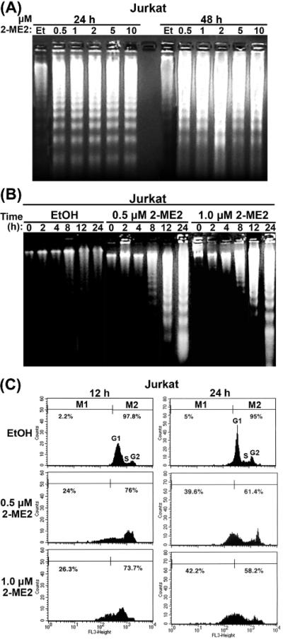
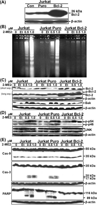
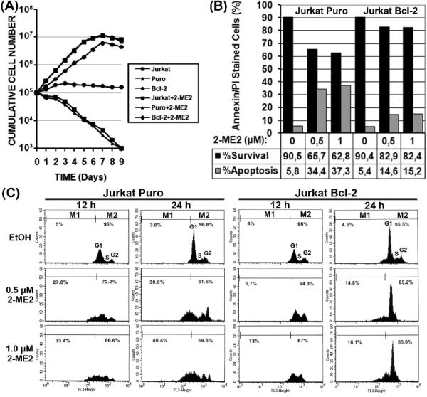
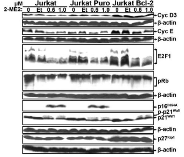
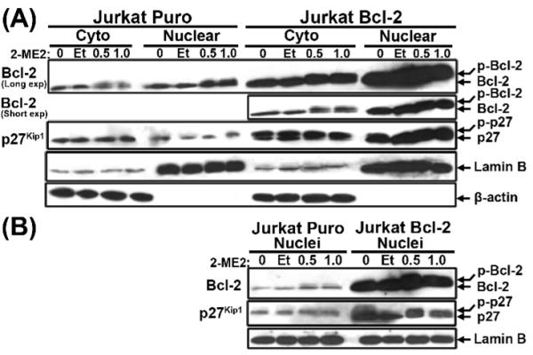
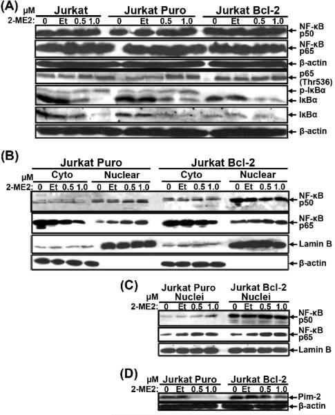
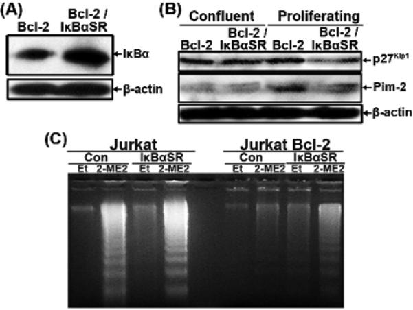
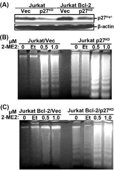
2-Methoxyestradiol (2-ME2) induces leukemia cells to undergo apoptosis in association with Bcl-2 inactivation but the mechanisms whereby Bcl-2 contributes to protection against programmed cell death in this context remain unclear. Here we showed that 2-ME2 inhibited the proliferation of Jurkat leukemia cells by markedly suppressing the levels of cyclins D3 and E, E2F1 and p21(Cip1/Waf1) and up-regulating p16(INK4A). Further, 2-ME2 induced apoptosis of Jurkat cells in association with down-regulation and phosphorylation of Bcl-2 (as mediated by JNK), up-regulation of Bak, activation of caspases-9 and -3 and PARP-1 cleavage. To determine the importance and mechanistic role of Bcl-2 in this process, we enforced its expression in Jurkat cells by retroviral transduction. Enforcing Bcl-2 expression in Jurkat cells abolished 2-ME2-induced apoptosis and instead produced a G1/S phase cell cycle arrest in association with markedly increased levels of p27(Kip1). Bcl-2 and p27(Kip1) were localized mainly in the nucleus in these apoptotic resistant cells. Interestingly, NF-kappaB activity and p50 levels were increased by 2-ME2 and suppression of NF-kappaB signaling reduced p27(Kip1) expression and sensitized cells to 2-ME2-induced apoptosis. Importantly, knocking-down p27(Kip1) in Jurkat Bcl-2 cells sensitized them to spontaneous and 2-ME2-induced apoptosis. Thus, Bcl-2 prevented the 2-ME2-induced apoptotic response by orchestrating a p27(Kip1)-dependent G1/S phase arrest in conjunction with activating NF-kappaB. Thus, we achieved a much better understanding of the penetrance and mechanistic complexity of Bcl-2 dependent anti-apoptotic pathways in cancer cells and why Bcl-2 inactivation is so critical for the efficacy of apoptosis and anti-proliferative inducing drugs like 2-ME2.


Figures

Fig. 1
Fig. 1
Induction of apoptosis of Jurkat cells by 2-ME2. (A) Dose-dependent induction of apoptosis of Jurkat cells. 1.5 × 10⁶ cells were treated with increasing concentrations of 2-ME2 for 24 and 48 h as indicated or ethanol (Et) as control and low molecular weight DNA was extracted and analyzed by agarose gel electrophoresis. (B) Time-dependent apoptosis of Jurkat cells. 1.5 × 10⁶ cells were treated with 0.5 and 1.0 μM of 2-ME2 or Et for different periods of time as indicated and low molecular weight DNA was extracted and analyzed by agarose gel electrophoresis. (C) Analysis of apoptosis of asynchronous cultures of Jurkat cells treated with 2-ME2 as determined by flow cytometry. Cells treated with 0.5 and 1.0 μM of 2-ME2 or Et for 12 and 24 h were subjected to flow cytometric analysis as described under materials and methods. Results were reproduced in two independent experiments.
Fig. 2
Fig. 2
Bcl-2 suppressed Jurkat cell apoptosis induced by 2-ME2 through the mitochondrial pathway. (A) Expression of Bcl-2 in retrovirally-transduced Jurkat cells. Total proteins extracted from Jurkat, Jurkat Puro and Jurkat Bcl-2 cells were analyzed by immunobloting for the expression of Bcl-2. (B) DNA fragmentation analysis of uninfected and retrovirus-infected Jurkat cells. DNA isolated from untreated and ethanol- or 2-ME2-treated Jurkat, Jurkat Puro and Jurkat Bcl-2 cells was analyzed by agarose gel electrophoresis. (C) Expression of Bcl-2 and Bak in control and Puro- or Bcl-2-expressing cells following treatment with 2-ME2. Total proteins isolated were analyzed by immunoblotting for the expression of Bcl-2- or Bak. In the Bcl-2 blot, the upper slower migrating band represents the phosphorylated form of Bcl-2. (D) Expression of total and phospho-JNK (Thr183/Tyr185) in control and Puro- or Bcl-2-expressing cells following treatment with 2-ME2. (E) Expression of caspases and PARP-1. Total proteins isolated as in (C) were analyzed for the expression of caspase-9, caspase-3 and PARP-1 or β-actin. All experiments were repeated 2-3 times with comparable results.
Fig. 3
Fig. 3
Bcl-2-mediated apoptotic block is linked to G1/S cell cycle arrest following treatment with 2-ME2. (A) Growth curves of uninfected and retrovirus-infected Jurkat cells in the presence or absence of 2-ME2, over a period of nine days. Each point represents the average of two wells in three independent experiments. (B) Flow cytometric analysis of asynchronous cultures of Puro- and Bcl-2-expressing Jurkat cells treated with 2-ME2. Jurkat Puro and Jurkat Bcl-2 cells treated with 2-ME2 or ethanol (Et) for 12 and 24 h were subjected to flow cytometric analysis. (C) Evaluation of apoptosis by annexin V/PI staining. Jurkat Puro and Jurkat Bcl-2 cells were treated with ethanol or 1.0 μM 2-ME2 for 24 h and subjected to annexin V/PI staining. Black bars represent viable cells and grey bars represent apoptotic cells.
Fig. 4
Fig. 4
Expression of cell cycle regulatory proteins in control and Puro- or Bcl-2-expressing Jurkat cells following treatment with 2-ME2. Cells were left untreated or treated with ethanol, or 0.5 μM and 1.0 μM 2-ME2 for 24 h. Total proteins isolated from untreated and ethanol- or 2-ME2-treated cells were analyzed by immunoblotting for the expression of cyclin D3, cyclin E, E2F1, pRb p16INK4A, p21Cip1/Waf1 and p27Kip1 or β-actin. Experiments were repeated 2-3 times with comparable results.
Fig. 5
Fig. 5
Subcellular expression of Bcl-2 and p27Kip1 in Puro- or Bcl-2-expressing Jurkat cells following 2-ME2 treatment. Cytoplasmic and nuclear fractions (A) or Nuclei (B) were isolated from Jurkat Puro and Jurkat Bcl-2 cells cultured in the absence or presence of ethanol or 2-ME2. The fractions were analyzed by immunoblotting for the expression of Bcl-2 or p27Kip1. Equal loading and verification of extracts was detected using an anti-Lamin B or anti-β-actin. No actin was detected in nuclear extracts or from isolated nuclei. All experiments were repeated multple times with similar results.
Fig. 6
Fig. 6
Expression of NF-κB and Pim-2 in Puro- and Bcl-2-expressing Jurkat cells following 2-ME2 treatment. Total proteins (A), Cytoplasmic and nuclear fractions (B) or Nuclei (C) isolated from untreated, ethanol- or 2-ME2-treated Jurkat Puro and Jurkat Bcl-2 cells were subjected to immunoblotting for the expression of the indicated proteins. (D) Expression of Pim-2, a NF-κB target protein. Total proteins isolated from untreated and ethanol- or 2-ME2-treated Jurkat Puro and Jurkat Bcl-2 cells were analyzed by immunoblotting for the expression of Pim-2. Equal loading and verification of extracts was detected using a rabbit polyclonal anti-Lamin B or a mouse monoclonal anti-β-actin. Experiments were repeated twice producing similar results.
Fig. 7
Fig. 7
Suppression of NF-κB signaling pathway in Jurkat cells. Jurkat and Jurkat Bcl-2 cells were infected with recombinant retrovirus carrying α IκBαSR to generate Jurkat IκBαSR and Jurkat Bcl-2/IκBαSR cells. (A) Total lysates from all the different cell types were analyzed by immunoblotting for the expression of IκBα or β-actin. (B) Total lysates from all the different cell types were analyzed by western immunoblot for the expression of p27Kip1 and Pim-2 or β-actin. (C) DNA was isolated from untreated, ethanol- or 2-ME2-treated cells as indicated and analyzed by agarose gel electrophoresis. Similar results were obtained in another independent experiment (data not shown.
Fig. 8
Fig. 8
Reduction of p27Kip1 levels sensitized Jurkat and Jurkat Bcl-2 cells to apoptosis. Jurkat and Jurkat Bcl-2 cells were infected with recombinant retrovirus bearing shRNA to p27Kip1 or a control vector to generate Jurkat Vec Jurkat p27KD, Jurkat Bcl-2/Vec and Jurkat Bcl-2/p27KD cells. (A) Total lysates from all the different cell types were analyzed by immunoblotting for the expression of p27Kip1 or β-actin. (B) and (C) Low molecular weight DNA was isolated from untreated, ethanol- or 2-ME2-treated Jurkat/Vec and Jurkat p27KD cells (B) or Jurkat Bcl-2/Vec and Jurkat Bcl-2/p27KD cells (C), as indicated and analyzed by agarose gel electrophoresis. Vec, vector pSR-Puro. All experiments were repeated twice with comparable results.

References

    1. Fotsis T, Zhang Y, Pepper MS, Adlercreutz H, Montesano R, Nawroth PP, et al. The endogenous estrogen metabolite 2-methoxyoestradiol inhibits angiogenesis and suppresses tumour growth. Nature. 1994;368:237–39. - PubMed
    1. D’Amato RJ, Lin CM, Flynn E, Folkman J, Hamel E. 2-methoxyestradiol, an endogenous mammalian metabolite, inhibits tubulin polymerization by interacting at the colchicine site. Proc Natl Acad Sci USA. 1994;91:3964–68. - PMC - PubMed
    1. Klauber N, Parangi S, Flynn E, Hamel E, D’Amato RJ. Inhibition of angiogenesis and breast cancer in mice by microtubule inhibitors 2-methoxyestradiol and taxol. Cancer Res. 1997;57:81–86. - PubMed
    1. Pribluda VS, Gubish ER, Jr, LaVallee TM, Treston A, Swartz GM, Green SJ. 2-methoxyestradiol: an endogenous antiangiogenic and antiproliferative drug candidate. Cancer Metastasis Rev. 2000;19:173–79. - PubMed
    1. Ryoo JJ, Cole CE, Anderson KC. Novel therapies for multiple myeloma. Blood Rev. 2002;16:167–74. - PubMed

Publication types

MeSH terms