. 2009 Aug;297(2):L219-27.
doi: 10.1152/ajplung.00043.2009. Epub 2009 May 15.

Claudin-4 augments alveolar epithelial barrier function and is induced in acute lung injury

Affiliations

Claudin-4 augments alveolar epithelial barrier function and is induced in acute lung injury

Charlie Wray et al. Am J Physiol Lung Cell Mol Physiol. 2009 Aug.

Abstract

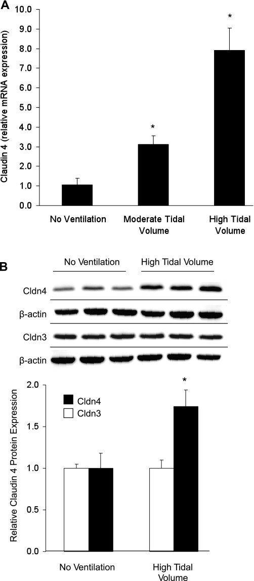
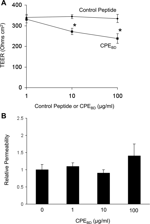
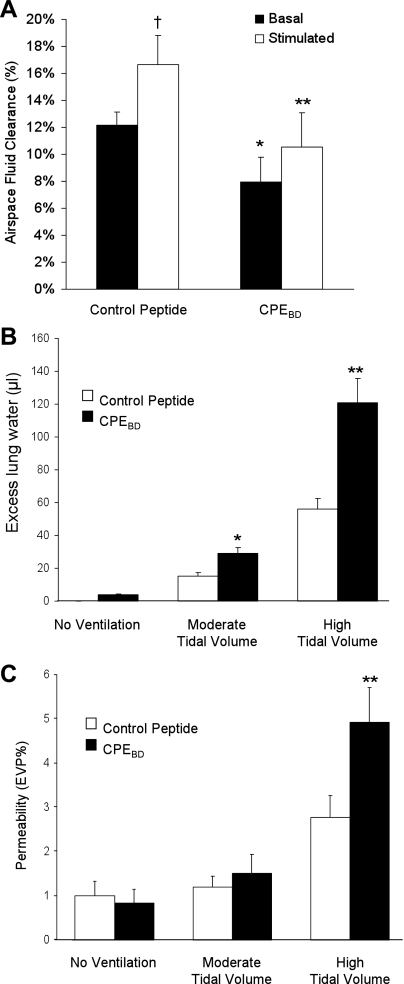
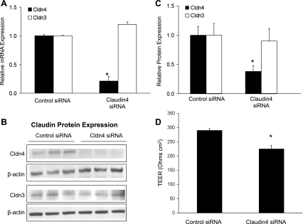
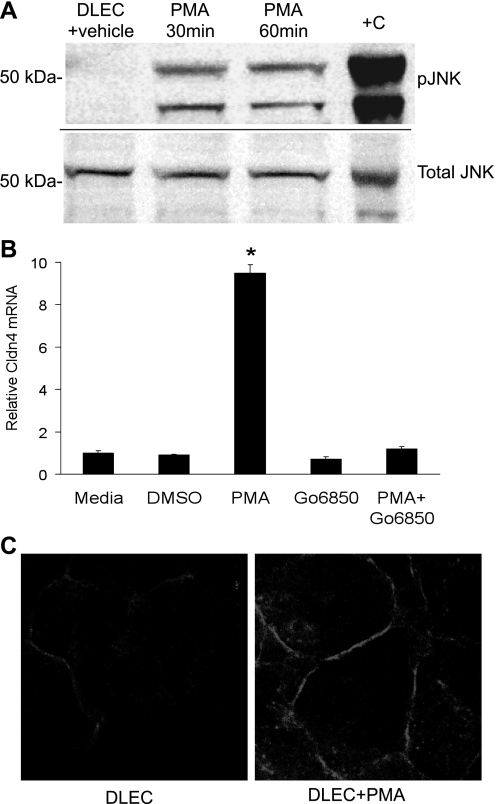
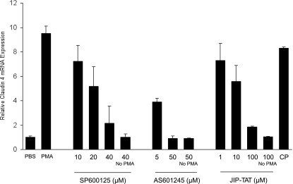
Intact alveolar barrier function is associated with better outcomes in acute lung injury patients; however, the regulation of alveolar epithelial paracellular transport during lung injury has not been extensively investigated. This study was undertaken to determine whether changes in tight junction claudin expression affect alveolar epithelial barrier properties and to determine the mechanisms of altered expression. In anesthetized mice exposed to ventilator-induced lung injury, claudin-4 was specifically induced among tight junction structural proteins. Real-time PCR showed an eightfold increase in claudin-4 expression in the lung injury model. To examine the role of this protein in barrier regulation, claudin-4 function was inhibited with small interfering RNA (siRNA) and a blocking peptide derived from the binding domain of Clostridium perfringens enterotoxin (CPE(BD)). Inhibition of claudin-4 decreased transepithelial electrical resistance but did not alter macromolecule permeability in primary rat and human epithelial cells. In mice, CPE(BD) decreased air space fluid clearance >33% and resulted in pulmonary edema during moderate tidal volume ventilation that did not induce edema in control peptide-treated mice. In vitro phorbol ester induced a ninefold increase in claudin-4 expression that was dependent on PKC activation and the JNK MAPK pathway. These data establish that changes in alveolar epithelial claudin expression influence paracellular transport, alveolar fluid clearance rates, and susceptibility to pulmonary edema. We hypothesize that increased claudin-4 expression early in acute lung injury represents a mechanism to limit pulmonary edema and that the regulation of alveolar epithelial claudin expression may be a novel target for acute lung injury therapy.


Figures

Fig. 1.
Fig. 1.
Claudin-4 (Cldn4) mRNA and protein expression in acute lung injury. A: real-time PCR demonstrated a dose-response increase in claudin-4 mRNA abundance in whole mouse lungs following 3 h of moderate or high tidal volume ventilation (normalized to β-actin) (*P < 0.05 compared with all other groups; n = 6 in each group). B: mechanical ventilation with high tidal volume for 3 h also significantly increased claudin-4 protein expression as shown by this Western blot. Densitometry revealed a significant 75% increase in claudin-4 protein and no change in claudin-3 protein expression with high tidal volume ventilation. Claudin-18 protein expression was also unchanged by mechanical ventilation (see text).
Fig. 2.
Fig. 2.
Blocking claudin-4 with a Clostridium perfringens binding domain peptide (CPEBD) alters paracellular permeability without dissociating tight junctions. A: CPEBD did not affect occludin (green) distribution but decreased claudin-4 (red) abundance at tight junctions in primary rat alveolar type II cells. The merged images on the right show the colocalization of claudin-4 and occludin at cell-cell junctions (yellow) in the control peptide-treated cells. B: claudin-18 expression was also not changed by CPEBD, however, CPEBD decreased claudin-3 expression. These data indicate that CPEBD results in a decrease in claudin-4 at tight junctions but does not entirely disrupt the tight junction. C: CPEBD also decreased claudin-4 protein levels in mouse whole lung without mechanical ventilation (*P < 0.05) and after 3 h of high tidal volume (HVT) ventilation (n = 3 per group; **P < 0.05). CPEBD decreased claudin-3 expression but did not affect claudin-18 protein expression in whole lungs (see text).
Fig. 3.
Fig. 3.
Effect of CPEBD on transepithelial electrical resistance (TEER) and permeability in vitro. A: CPEBD decreased transepithelial electrical resistance in a dose-dependent fashion in primary rat type II epithelial cells (means ± SE; *P < 0.05 compared with control peptide; n = minimum of 3 replicates of 6 wells each). B: CPEBD did not increase paracellular permeability to large (40-kDa) molecules indicating that paracellular transport properties were altered, but tight junction function was not lost (*P < 0.05 compared with control peptide; n = minimum of 3 replicates of 6 wells each, means ± SE).
Fig. 4.
Fig. 4.
Effect of the loss of claudin-4 on air space fluid clearance, pulmonary edema, and albumin flux in vivo. A: blocking claudin-4 with CPEBD in vivo decreased basal and β-adrenergic-stimulated air space fluid clearance. CPEBD decreased basal air space fluid transport (% instilled volume cleared in 30 min) in unventilated mice (black bars). CPEBD also decreased β-adrenergic agonist-stimulated (terbutaline, 10−5 M) alveolar fluid clearance (white bars) (*P < 0.05 compared with control peptide basal clearance and **P < 0.05 compared with control peptide-stimulated clearance; n = 6–9 in each group). β-Adrenergic agonist increased fluid clearance in control peptide-treated mice (†P < 0.05) but not in CPEBD-treated mice. B: blocking claudin-4 with CPEBD in vivo increases pulmonary edema during injury. CPEBD (black bars) resulted in increased pulmonary edema (excess lung water) during either moderate or high tidal volume mechanical ventilation. CPEBD did not significantly affect baseline lung water in the absence of mechanical ventilation. C: CPEBD did not affect permeability to albumin at baseline or during moderate tidal volume ventilation. However, CPEBD increased lung injury severity with high tidal volume mechanical ventilation as measured by albumin flux expressed as the percent of the plasma volume in the extravascular spaces of the lung [extravascular plasma equivalents (EVP%)]. *P < 0.05 compared with control peptide (white bars), **P < 0.05 compared with all other groups; n = 6–9 in each group. Therefore, inhibition of claudin-4 function with CPEBD decreased air space fluid clearance, increased susceptibility to pulmonary edema with mechanical ventilation, and resulted in more severe ventilator-induced lung injury.
Fig. 5.
Fig. 5.
Claudin-4 knockdown with small interfering RNA (siRNA). To extend and confirm the CPEBD studies by specifically blocking only claudin-4 expression, primary human type II-like distal lung epithelial cells (DLECs) were transfected with siRNA targeted to claudin-4. A: at 72 h posttransfection, claudin-4 siRNA (20 nM) decreased claudin-4 mRNA abundance without affecting claudin-3 expression (*P < 0.05 compared with nonsilencing control siRNA without homology to any known human gene; n = minimum of 3 replicates of 6 wells each, means ± SE). B: claudin-4 siRNA significantly decreased claudin-4 but not claudin-3 protein expression. C: densitometry of Western blot data normalized to β-actin showed a significant decrease in claudin-4 protein with targeted siRNA (n = 3 per group; P < 0.05; means ± SD) but no change in claudin-3 protein levels. D: claudin-4 knockdown decreased transepithelial electrical resistance in human cells with an effect size comparable with CPEBD (*P < 0.05 compared with nonsilencing control siRNA; n = minimum of 3 replicates of 6 wells each, means ± SE). These data support the hypothesis that claudin-4 expression increases transepithelial resistance and decreases paracellular ion transport.
Fig. 6.
Fig. 6.
PMA induces the JNK MAPK pathway and claudin-4 expression. A: in human type II-like DLECs, PMA-induced JNK1 (46 kDa) and JNK2 (54 kDa) phosphorylation (pJNK) at 30 and 60 min compared with vehicle (DMSO). UV-treated 293 cell extracts (Cell Signaling) were used as a positive control (+C). The same samples were tested for phosphorylated JNK and total JNK on separate, identically loaded polyacrylamide gels run concurrently. B: PMA induced a 9-fold increase in claudin-4 mRNA expression at 4 h that was completely inhibited by the classic and novel PKC inhibitor Gö-6850 (1 μM; *P < 0.05; n = 4 replicates of 6 wells each, means ± SE). C: PMA increased claudin-4 protein abundance primarily at the plasma membrane at 4 h.
Fig. 7.
Fig. 7.
PMA-mediated claudin-4 induction requires the JNK pathway. A: inhibition of the JNK MAPK pathway with each of 3 inhibitors (SP-600125, AS601245, and JIP-TAT peptide) blocked the PMA-induced increase in claudin-4 expression in human DLECs in a dose-dependent fashion (*P < 0.05; n = minimum of 3 replicates of 6 wells each, means ± SE). CP, control peptide TAT. Inhibitors of ERK1/2 or p38 had no effect on PMA-mediated claudin-4 mRNA expression.

Comment in

References

    1. Adir Y, Factor P, Dumasius V, Ridge KM, Sznajder JI. Na,K-ATPase gene transfer increases liquid clearance during ventilation-induced lung injury. Am J Respir Crit Care Med 168: 1445–1448, 2003. - PubMed
    1. Borok Z, Hami A, Danto SI, Lubman RL, Kim KJ, Crandall ED. Effects of EGF on alveolar epithelial junctional permeability and active sodium transport. Am J Physiol Lung Cell Mol Physiol 270: L559–L565, 1996. - PubMed
    1. Chen SP, Zhou B, Willis BC, Sandoval AJ, Liebler JM, Kim KJ, Ann DK, Crandall ED, Borok Z. Effects of transdifferentiation and EGF on claudin isoform expression in alveolar epithelial cells. J Appl Physiol 98: 322–328, 2005. - PubMed
    1. Coyne CB, Gambling TM, Boucher RC, Carson JL, Johnson LG. Role of claudin interactions in airway tight junctional permeability. Am J Physiol Lung Cell Mol Physiol 285: L1166–L1178, 2003. - PubMed
    1. Coyne CB, Vanhook MK, Gambling TM, Carson JL, Boucher RC, Johnson LG. Regulation of airway tight junctions by proinflammatory cytokines. Mol Biol Cell 13: 3218–3234, 2002. - PMC - PubMed

Publication types

MeSH terms