. 2009 Jul;150(3):1260-71.
doi: 10.1104/pp.109.136812. Epub 2009 May 15.

LPA66 is required for editing psbF chloroplast transcripts in Arabidopsis

Affiliations

LPA66 is required for editing psbF chloroplast transcripts in Arabidopsis

Wenhe Cai et al. Plant Physiol. 2009 Jul.

Erratum in

  • CORRECTION: Vol. 150: 1260-1271, 2009.
    [No authors listed] [No authors listed] Plant Physiol. 2017 Feb;173(2):1522-1523. doi: 10.1104/pp.16.01911. Plant Physiol. 2017. PMID: 28154116 Free PMC article. No abstract available.

Abstract

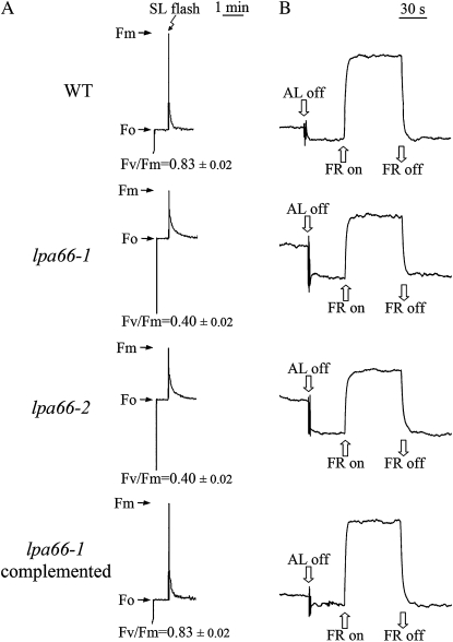
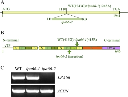
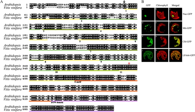
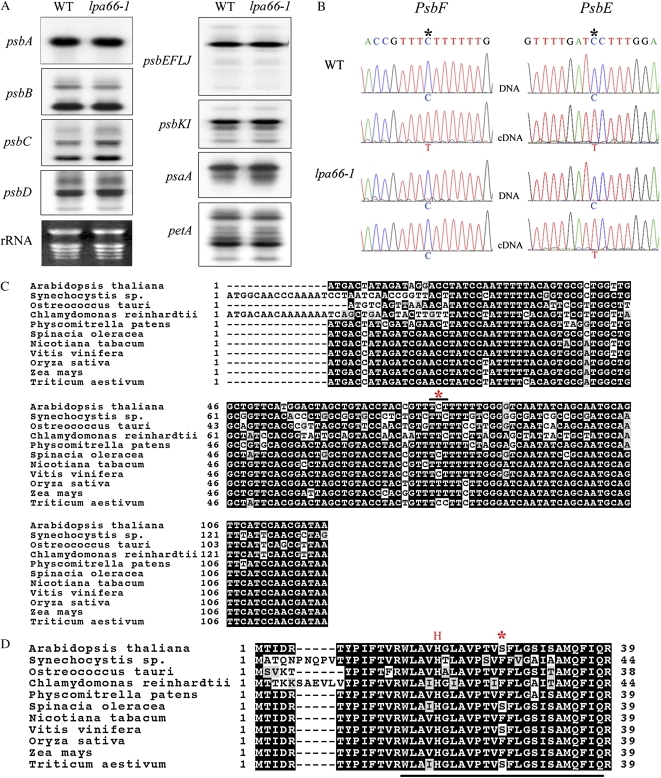
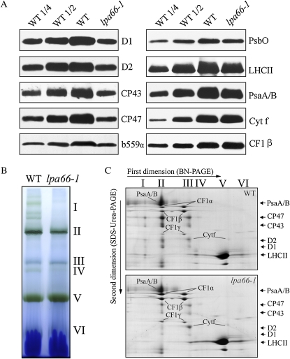
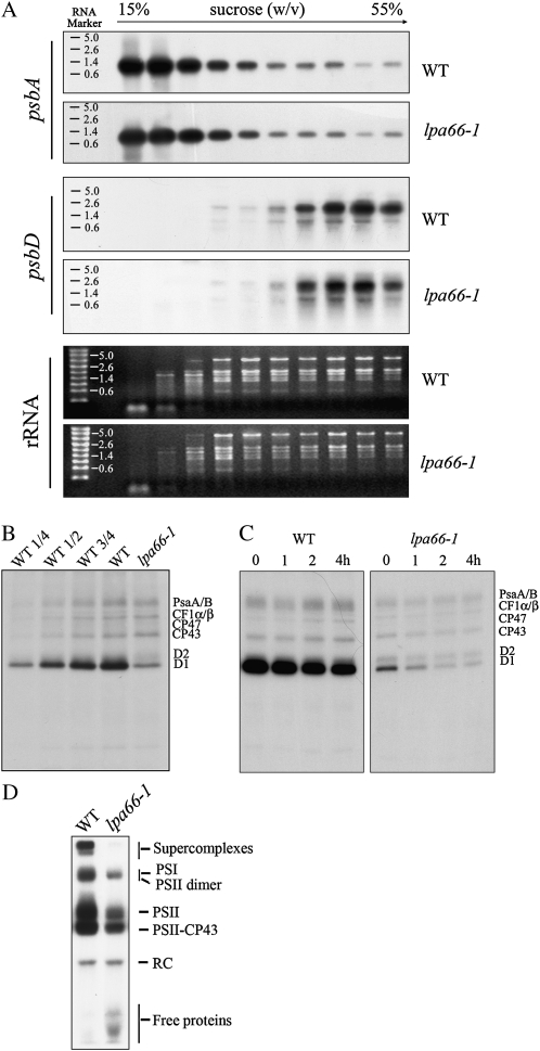
To gain insight into the molecular mechanism of RNA editing, we have characterized the low psii accumulation66 (lpa66) Arabidopsis (Arabidopsis thaliana) mutant, which displays a high chlorophyll fluorescence phenotype. Its perturbed chlorophyll fluorescence is reflected in reduced levels of photosystem II (PSII) proteins. In vivo protein labeling showed that synthesis rates of the PSII reaction center protein D1/D2 were lower, and turnover rates of PSII core proteins higher, than in wild-type counterparts. The assembly of newly synthesized proteins into PSII occurs in the lpa66 mutant but with reduced efficiency compared with the wild type. LPA66 encodes a chloroplast protein of the pentatricopeptide repeat family. In lpa66 mutants, editing of psbF that converts serine to phenylalanine is specifically impaired. Thus, LPA66 is specifically required for editing the psbF transcripts in Arabidopsis, and the amino acid alternation due to lack of editing strongly affects the efficiency of the assembly of PSII complexes.


Figures

Figure 1.
Figure 1.
Spectroscopic analysis of wild-type (WT), lpa66-1 and lpa66-2 mutant, and lpa66-1 complemented transgenic plants. A, Chlorophyll fluorescence induction. Fm, Maximum fluorescence yield; Fo, minimum fluorescence yield when PSII centers are open; SL, saturating light. The ratios of variable to maximum fluorescence, reflecting the maximum potential of PSII photochemical reactions, were calculated from Fv/Fm = (FmFo)/Fm. B, Redox kinetics of P700. The oxidation state of P700 was investigated by measuring absorbance changes of P700 at 820 nm induced by far-red light (FR; 720 nm).
Figure 2.
Figure 2.
Identification of the lpa66 mutation. A, Schematic diagram of the LPA66 gene. The box (1–1,941bp) represents the coding region of LPA66 from ATG to TGA without introns. The lpa66-1 and lpa66-2 mutations are indicated. LB and RB represent the left and right borders, respectively, of the inserted T-DNA in the lpa66-2 line. The diagram is not drawn to scale. B, Predicted motif structure of LPA66. The designations of the P, L, L2, S, C-terminal E motif, and DYW domain correspond to those proposed by Lurin et al. (2004). cTP indicates the plastid transit peptide. The start and end positions of LPA66 are indicated as 1 and 646, respectively. C, RT-PCR analysis of mutant plants. RT-PCR was performed with specific primers for LPA66 and actin genes. WT, Wild type.
Figure 3.
Figure 3.
LPA66 sequence alignment and subcellular localization. A, Amino acid alignment of LPA66 (At5g48910). The amino acid sequence of At5g48910 was compared with the homologous sequence from grapevine (LOC100261359). The predicted cleavage site for the plastid transit peptide is indicated by an arrow. PPR motifs are numbered. The orange line beneath the sequences indicates the E motif. The DYW domain is purple underlined, and the mutated amino acid in lpa66 is marked by the red asterisk. B, Subcellular localization of the LPA66 protein according to fluorescence signals visualized using confocal laser scanning microscopy. The indicated fusion proteins with sGFP were transiently expressed in protoplasts under the control of the cauliflower mosaic virus 35S promoter, and the green GFP signals were obtained by confocal microscopy (A, D, G, and J). The chloroplasts were visualized by chlorophyll autofluorescence (B, E, H, and K). The colocalization of GFP and chloroplasts is indicated in merged images (C, F, I, and L). The constructs used for transformation are indicated to the right: Nuc-GFP, control with nuclear localization signal of fibrillarin; Mit-GFP, control with mitochondrial localization signal of FRO1; Chl-GFP, control with the transit peptide of the ribulose bisphosphate carboxylase small subunit; LPA66-GFP, signals from the LPA66-GFP fusion protein.
Figure 4.
Figure 4.
RNA transcripts and editing analysis in lpa66 mutants. A, RNA gel blot hybridization with total RNA from leaves of wild-type (WT) and lpa66-1 mutant plants. Ten micrograms of total leaf RNA per well from 5-week-old wild-type and lpa66-1 plants was loaded per well. The probes for the genes psbA, psbB, psbC, psbD, psbEFLJ, psbKI, psaA, and petA are indicated to the left. rRNA was visualized by staining with ethidium bromide as an equal loading control. B, Analysis of RNA editing of psbF and psbE transcripts. RT-PCR products containing the psbF and psbE editing sites (asterisks) were directly sequenced. C and D, Alignments of psbF gene sequences (C) and predicted proteins directly translated from genomic sequences (D) from various organisms. The chloroplast genomic accession numbers are NC_000932 (Arabidopsis thaliana), NC_000911 (Synechocystis PCC 6803), NC_008289 (Ostreococcus tauri), NC_005353 (Chlamydomonas reinhardtii), NC_005087 (Physcomitrella patens), NC_002202 (Spinacia oleracea), NC_001879 (Nicotiana tabacum), DQ424856 (Vitis vinifera), NC_001320 (Oryza sativa), NC_001666 (Zea mays), and NC_002762 (Triticum aestivum). The editing site and its corresponding amino acid residue are indicated by asterisks (C and D, respectively); the His residue for heme binding is indicated by the red H, and the transmembrane region is underlined in D.
Figure 5.
Figure 5.
Levels of chloroplast proteins. A, Immunodetection of chloroplast proteins. The total protein extracts (10 μg of proteins) from 5-week-old wild-type (WT) and lpa66-1 leaves were separated by SDS-urea-PAGE and blotted, and the blots were probed with specific antibodies: anti-D1, anti-D2, anti-CP43, anti-CP47, anti-cytochrome b559α, anti-PsbO, anti-LHCII, anti-PsaA/B, anti-cytochrome f, and anti-CF1β. B, BN gel analysis of thylakoid membrane protein complexes. Thylakoid membranes (10 μg of chlorophyll) from 5-week-old wild-type and lpa66-1 leaves were solubilized with 1% dodecyl-β-d-maltoside and separated by 6% to 12% gradient BN gel electrophoresis. The positions of protein complexes representing PSII supercomplexes (I), monomeric PSI and dimeric PSII (II), monomeric PSII (III), CP43 minus PSII (IV), trimeric LHCII/PSII reaction center (V), and monomeric LHCII (VI) are identified as described previously (Guo et al., 2005; Peng et al., 2006). C, Two-dimensional separation of protein complexes in the thylakoid membranes. After separated on the BN gel, the complex proteins in a single lane were further separated by 15% SDS-urea-PAGE and stained with Coomassie Brilliant Blue. Names of the proteins resolved by the second-dimension SDS-PAGE, previously identified, are indicated by arrows (Peng et al., 2006).
Figure 6.
Figure 6.
Polysome association and in vivo labeling of wild-type (WT) and lpa66-1 plants. A, Association of psbA and psbD mRNAs with polysomes. Ten fractions of equal volume were collected from the top to bottom of 15% to 55% Suc gradients, and equal proportions of the RNA purified from each fraction were analyzed by gel-blot hybridization. rRNAs were detected by ethidium bromide staining. The RNA size markers are indicated to the left. B, Pulse labeling of thylakoid membrane proteins. After pulse labeling young Arabidopsis seedlings in the presence of cycloheximide for 20 min, thylakoid membranes were isolated, and the proteins were separated by SDS-urea-PAGE and visualized autoradiographically. C, Pulse and chase labeling of thylakoid membrane proteins. After pulse labeling for 20 min followed by 1-, 2-, or 4-h chases with cold Met, thylakoid membranes were isolated, and the proteins were separated by SDS-urea-PAGE and visualized autoradiographically. D, BN gel analysis of labeled thylakoid membrane protein complexes after pulse labeling. After a 20-min pulse in Arabidopsis young seedlings in the presence of cycloheximide, the thylakoid membranes were isolated and solubilized with dodecyl-β-d-maltoside, then the protein complexes were separated by BN-PAGE and visualized autoradiographically. Bands corresponding to various PSII assembly complexes are indicated to the right.

Similar articles

Cited by

References

    1. Andrés C, Lurin C, Small ID (2007) The multifarious roles of PPR proteins in plant mitochondrial gene expression. Physiol Plant 129 14–22
    1. Barkan A (1988) Proteins encoded by a complex chloroplast transcription unit are each translated from both monocistronic and polycistronic mRNAs. EMBO J 7 2637–2644 - PMC - PubMed
    1. Barkan A, Goldschmidt-Clermont M (2000) Participation of nuclear genes in chloroplast gene expression. Biochimie 82 559–572 - PubMed
    1. Beick S, Schmitz-Linneweber C, Williams-Carrier R, Jensen B, Barkan A (2008) The pentatricopeptide repeat protein PPR5 stabilizes a specific tRNA precursor in maize chloroplasts. Mol Cell Biol 28 5337–5347 - PMC - PubMed
    1. Bock R (2000) Sense from nonsense: how the genetic information of chloroplasts is altered by RNA editing. Biochimie 82 549–557 - PubMed

Publication types

Associated data

LinkOut - more resources