Review
. 2009 Jun;212(Pt 11):1611-9.
doi: 10.1242/jeb.030007.

Vacuolar-type proton pumps in insect epithelia

Affiliations
Review

Vacuolar-type proton pumps in insect epithelia

Helmut Wieczorek et al. J Exp Biol. 2009 Jun.

Abstract

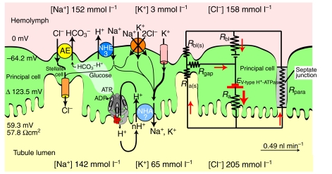
Active transepithelial cation transport in insects was initially discovered in Malpighian tubules, and was subsequently also found in other epithelia such as salivary glands, labial glands, midgut and sensory sensilla. Today it appears to be established that the cation pump is a two-component system of a H(+)-transporting V-ATPase and a cation/nH(+) antiporter. After tracing the discovery of the V-ATPase as the energizer of K(+)/nH(+) antiport in the larval midgut of the tobacco hornworm Manduca sexta we show that research on the tobacco hornworm V-ATPase delivered important findings that emerged to be of general significance for our knowledge of V-ATPases, which are ubiquitous and highly conserved proton pumps. We then discuss the V-ATPase in Malpighian tubules of the fruitfly Drosophila melanogaster where the potential of post-genomic biology has been impressively illustrated. Finally we review an integrated physiological approach in Malpighian tubules of the yellow fever mosquito Aedes aegypti which shows that the V-ATPase delivers the energy for both transcellular and paracellular ion transport.


Figures

Fig. 1.
Fig. 1.
Cryosection of the tobacco hornworm midgut epithelium. (A) Immunolocalization of the V1 subunit A by a monoclonal antibody. (B) Same section as in A, but visualized by Hoffman modulation contrast microscopy. a, apical side; b, basal side; BB, brush border the columnar cells; GC goblet cavity of goblet cells. The V-ATPase is present only in the apical membrane of goblet cells.
Fig. 2.
Fig. 2.
Model of a eukaryotic V-ATPase. (A) The peripheral V1 complex consists of eight different subunits identified with capital letters A–H. The integral membrane VO complex consists of at least four different subunits identified with lowercase letters (a, c, d, e). In yeast subunit c occurs together with its isoforms c′ and c″. Actin filaments bind to subunits B (Holliday et al., 2000) and C (Vitavska et al., 2003). (B) Disassembly of the V-ATPase into its V1 and VO complexes. Subunit C, which dissociates from the V1 complex, binds not only to F actin but also to monomeric G-actin (Vitavska et al., 2005).
Fig. 3.
Fig. 3.
Phosphorylation of the V1 subunit C by protein kinase A (PKA) and its assumed involvement in the process of reversible V1VO disassembly.
Fig. 4.
Fig. 4.
The V-ATPase in Malpighian tubules of the mosquito Aedes aegypti. (A) Cross-section of the abdomen. Immunofluorescence labeling of subunit C of the V-type H+-ATPase at the luminal brush border of Malpighian tubules (courtesy of H. Merzendorfer). (B) Sections of Malpighian tubules. Immunoperoxidase labeling of the B-subunit of the V-type H+-ATPase at the brush border of principal cells. The V-ATPase is not present in stellate cells (adapted from Weng et al., 2003). Scale bars in A and B are 100 μm.
Fig. 5.
Fig. 5.
Molecular and electrical models for powering the transepithelial secretion of NaCl, KCl and water in Malpighian tubules of Ae. aegypti under control conditions. Principal cells mediate the transepithelial active transport of Na+ and K+ driven by the V-type H+ ATPase collaborating with a putative electrogenic H+/cation exchanger (NHA?) located at the apical membrane. Stellate cells and the septate junction mediate the passive transepithelial secretion of Cl. Active and passive transepithelial transport routes are electrically coupled via septate and gap junctions. Red arrows indicate the flow of positive charge, where current can be carried by negative charge (anion) flowing in the opposite direction. R, resistance; a and a(s) apical membrane of principal and stellate cell, respectively; bl and bl(s), basolateral membrane of principal and stellate cell, respectively; gap, gap junction; para, paracellular pathway; AE, electroneutral anion exchanger; NHE3, electroneutral Na+/H+ exchanger type 3. The paracellular pathway between principal and stellate cells is not depicted. Data from previous publications (Beyenbach, 2001; Beyenbach and Masia, 2002; O'Connor and Beyenbach, 2001; Piermarini et al., 2008).

Similar articles

Cited by

References

    1. Al Awqati, Q. (1978). H+ transport in urinary epithelia. Am. J. Physiol. 235, F77-F88. - PubMed
    1. Allan, A. K., Du, J., Davies, S. A. and Dow, J. A. (2005). Genome-wide survey of V-ATPase genes in Drosophila reveals a conserved renal phenotype for lethal alleles. Physiol. Genomics 22, 128-138. - PubMed
    1. Azuma, M., Harvey, W. R. and Wieczorek, H. (1995). Stoichiometry of K+/H+ antiport helps to explain extracellular pH 11 in a model epithelium. FEBS Lett. 361, 153-156. - PubMed
    1. Bertram, G. and Wessing, A. (1994). Intracellular pH regulation by the plasma membrane V-ATPase in Malpighian tubules of Drosophila larvae. J. Comp. Physiol. B 164, 238-246. - PubMed
    1. Bertram, G., Schleithoff, L., Zimmermann, P. and Wessing, A. (1991). Bafilomycin A1 is a potent inhibitor of urine formation by Malpighian tubules of Drosophila hydei: is a vacuolar-type ATPase involved in ion and fluid secretion? J. Insect Physiol. 37, 201-209.

Publication types

MeSH terms

Substances

LinkOut - more resources