

Carbonic anhydrases and anion transport in mosquito midgut pH regulation
- PMID: 19448076
- PMCID: PMC2683011
- DOI: 10.1242/jeb.028084
Carbonic anhydrases and anion transport in mosquito midgut pH regulation
Abstract
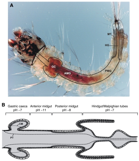
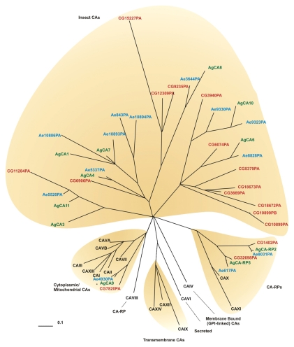
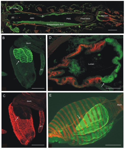
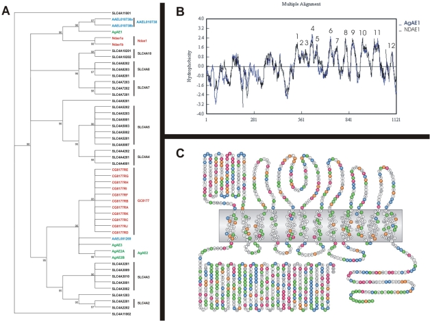
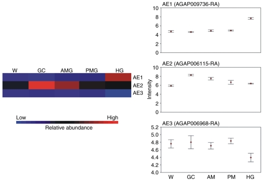
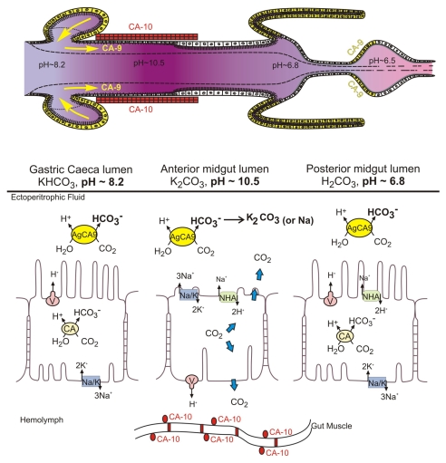
Mosquito larvae use a digestive strategy that is relatively rare in nature. The anterior half of the larval mosquito midgut has a luminal pH that ranges between 10.5 and 11.5. Most other organisms, both large and small, initiate digestion in an acid medium. The relative uniqueness of the highly alkaline digestive strategy has been a long-standing research focus in larval lepidopterans. More recently, the disease vector potential of mosquitoes has fueled specific interest in larval mosquito biology and the alkaline digestive environment in the midgut. The probable principle anion influencing the highly alkaline gut lumen is bicarbonate/carbonate. Bicarbonate/carbonate is regulated at least in part by the activity of carbonic anhydrases. Hence, we have focused attention on the carbonic anhydrases of the mosquito larva. Anopheles gambiae, the major malaria mosquito of Africa, is an organism with a published genome which has facilitated molecular analyses of the 12 carbonic anhydrase genes annotated for this mosquito. Microarray expression analyses, tissue-specific quantitative RT-PCR, and antibody localization have been used to generate a picture of carbonic anhydrase distribution in the larval mosquito. Cytoplasmic, GPI-linked extracellular membrane-bound and soluble extracellular carbonic anhydrases have been located in the midgut and hindgut. The distribution of the enzymes is consistent with an anion regulatory system in which carbonic anhydrases provide a continuous source of bicarbonate/carbonate from the intracellular compartments of certain epithelial cells to the ectoperitrophic space between the epithelial cells and the acellular membrane separating the food bolus from the gut cells and finally into the gut lumen. Carbonic anhydrase in specialized cells of the hindgut (rectum) probably plays a final role in excretion of bicarbonate/carbonate into the aquatic environment of the larva. Detection and characterization of classic anion exchangers of the SLC4A family in the midgut has been problematic. The distribution of carbonic anhydrases in the system may obviate the requirement for such transporters, making the system more dependent on simple carbon dioxide diffusion and ionization via the activity of the enzyme.
Figures
Similar articles
-
Characterization of Carbonic Anhydrase 9 in the Alimentary Canal of Aedes aegypti and Its Relationship to Homologous Mosquito Carbonic Anhydrases.Int J Environ Res Public Health. 2017 Feb 21;14(2):213. doi: 10.3390/ijerph14020213. Int J Environ Res Public Health. 2017. PMID: 28230813 Free PMC article.
-
A GPI-linked carbonic anhydrase expressed in the larval mosquito midgut.J Exp Biol. 2004 Dec;207(Pt 26):4559-72. doi: 10.1242/jeb.01287. J Exp Biol. 2004. PMID: 15579552
-
Cloning and characterization of AgCA9, a novel alpha-carbonic anhydrase from Anopheles gambiae Giles sensu stricto (Diptera: Culicidae) larvae.J Exp Biol. 2007 Nov;210(Pt 22):3919-30. doi: 10.1242/jeb.008342. J Exp Biol. 2007. PMID: 17981859
-
The Fundamental Role of Bicarbonate Transporters and Associated Carbonic Anhydrase Enzymes in Maintaining Ion and pH Homeostasis in Non-Secretory Organs.Int J Mol Sci. 2020 Jan 4;21(1):339. doi: 10.3390/ijms21010339. Int J Mol Sci. 2020. PMID: 31947992 Free PMC article. Review.
-
Epithelial ultrastructure and cellular mechanisms of acid and base transport in the Drosophila midgut.J Exp Biol. 2009 Jun;212(Pt 11):1731-44. doi: 10.1242/jeb.029306. J Exp Biol. 2009. PMID: 19448082 Review.
Cited by
-
Molecular and biochemical analysis of the beta class carbonic anhydrases in Caenorhabditis elegans.Mol Biol Rep. 2010 Jul;37(6):2941-50. doi: 10.1007/s11033-009-9857-z. Epub 2009 Oct 9. Mol Biol Rep. 2010. PMID: 19816790
-
Draft genome sequences of Hirudo medicinalis and salivary transcriptome of three closely related medicinal leeches.BMC Genomics. 2020 Apr 29;21(1):331. doi: 10.1186/s12864-020-6748-0. BMC Genomics. 2020. PMID: 32349672 Free PMC article.
-
Molecular basis of essential amino acid transport from studies of insect nutrient amino acid transporters of the SLC6 family (NAT-SLC6).J Insect Physiol. 2012 Apr;58(4):433-49. doi: 10.1016/j.jinsphys.2011.12.018. Epub 2012 Jan 2. J Insect Physiol. 2012. PMID: 22230793 Free PMC article. Review.
-
Biochemical and Functional Characterization of Glycoside Hydrolase Family 16 Genes in Aedes aegypti Larvae: Identification of the Major Digestive β-1,3-Glucanase.Front Physiol. 2019 Feb 28;10:122. doi: 10.3389/fphys.2019.00122. eCollection 2019. Front Physiol. 2019. PMID: 30873040 Free PMC article.
-
Dynamic gut microbiome across life history of the malaria mosquito Anopheles gambiae in Kenya.PLoS One. 2011;6(9):e24767. doi: 10.1371/journal.pone.0024767. Epub 2011 Sep 21. PLoS One. 2011. PMID: 21957459 Free PMC article.
References
-
- Black, W. C. and Kondratieff, B. C. (2005). Evolution of arthropod disease vectors. In Biology of Disease Vectors, 2nd edn (ed. W. C. Marquardt), pp. 9-23. Burlington, MA: Elsevier.
-
- Bradley, T. J. (1987). Physiology of osmoregulation in mosquitoes. Annu. Rev. Entomol. 32, 439-462. - PubMed
-
- Clark, T. M., Vieira, M. A., Huegel, K. L., Flury, D. and Carper, M. (2007). Strategies for regulation of hemolymph pH in acidic and alkaline water by the larval mosquito Aedes aegypti (L.) (Diptera;Culicidae). J. Exp. Biol. 210, 4359-4367. - PubMed
-
- Clements, A. N. (1992). The Biology of Mosquitoes, vol. 1 (ed. A. N. Clements). London: Chapman Hall.
Publication types
MeSH terms
Substances
Grants and funding
LinkOut - more resources
Full Text Sources
Medical
Molecular Biology Databases
