Review
. 2009 Jun;212(Pt 11):1762-72.
doi: 10.1242/jeb.028803.

Regulation of the V-ATPase in kidney epithelial cells: dual role in acid-base homeostasis and vesicle trafficking

Affiliations
Review

Regulation of the V-ATPase in kidney epithelial cells: dual role in acid-base homeostasis and vesicle trafficking

Dennis Brown et al. J Exp Biol. 2009 Jun.

Abstract

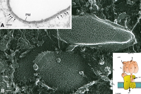
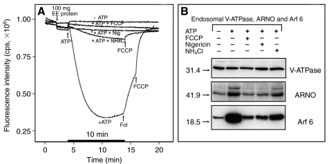
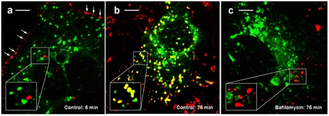
The proton-pumping V-ATPase is a complex, multi-subunit enzyme that is highly expressed in the plasma membranes of some epithelial cells in the kidney, including collecting duct intercalated cells. It is also located on the limiting membranes of intracellular organelles in the degradative and secretory pathways of all cells. Different isoforms of some V-ATPase subunits are involved in the targeting of the proton pump to its various intracellular locations, where it functions in transporting protons out of the cell across the plasma membrane or acidifying intracellular compartments. The former process plays a critical role in proton secretion by the kidney and regulates systemic acid-base status whereas the latter process is central to intracellular vesicle trafficking, membrane recycling and the degradative pathway in cells. We will focus our discussion on two cell types in the kidney: (1) intercalated cells, in which proton secretion is controlled by shuttling V-ATPase complexes back and forth between the plasma membrane and highly-specialized intracellular vesicles, and (2) proximal tubule cells, in which the endocytotic pathway that retrieves proteins from the glomerular ultrafiltrate requires V-ATPase-dependent acidification of post-endocytotic vesicles. The regulation of both of these activities depends upon the ability of cells to monitor the pH and/or bicarbonate content of their extracellular environment and intracellular compartments. Recent information about these pH-sensing mechanisms, which include the role of the V-ATPase itself as a pH sensor and the soluble adenylyl cyclase as a bicarbonate sensor, will be addressed in this review.


Figures

Fig. 1.
Fig. 1.
Structure of the V-ATPase. The large cytoplasmic oriented V1 sector of the V-ATPase holoenzyme can be visualized by conventional (A) and rapid-freeze, deep-etch (B) electron microscopy (EM). By conventional EM, the V-ATPase appears as dense projections (arrows) attached to the cytoplasmic side of the apical plasma membrane (PM) of this A-type intercalated cell (A-IC) from a rat kidney. Note that almost all of the membrane in this section is coated with the V-ATPase. Panel B shows the underside of a mitochondria-rich (MR)-cell from toad urinary bladder, coated with arrays of stud-like projections that correspond to V-ATPase V1 sectors and to the projections seen by thin section EM in panel A. Each projection is about 10 nm in diameter. The bottom right inset shows a schematic representation of the subunits that comprise the transmembrane Vo sector and the cytoplasmic V1 sector of the holoenzyme. See text for more details. Scale bar=50 nm.
Fig. 2.
Fig. 2.
Cryostat section of cortical collecting duct from a PLP (paraformaldehyde, lysine, periodate)-fixed rat kidney immunostained to reveal aquaporin 2 (AQP2) (green: anti-AQP2 C-terminus, raised in goat, followed by donkey anti-goat IgG coupled to Alexa 488) and the V-ATPase (red: anti-V-ATPase A-subunit C-terminus, raised in rabbit, followed by donkey anti-rabbit IgG coupled to CY3). Principal cells contain a tight apical band of AQP2 in this tissue and also show a weaker staining at their basolateral pole. Intercalated cells (IC) have either a strong apical staining only [A-type intercalated cells (A-IC)] or a basolateral/bipolar staining for the V-ATPase [B-type intercalated cells (B-IC)]. Nuclei are stained blue with DAPI. Scale bar=5 μm.
Fig. 3.
Fig. 3.
A-type intercalated cells (A-IC) show a very high rate of apical endocytosis. The cells in panel (A) are from a rat that was injected with 25 mg of FITC-dextran in 1 ml PBS as an endocytotic marker. Cells in this cryostat section of a PLP (paraformaldehyde, lysine, periodate)-fixed medullary collecting duct are identified as A-IC by the basolateral immunostaining for the anion exchanger AE1 (orange color). Anti-AE1 antibodies raised in rabbit were a gift from Dr Seth Alper, Beth Israel Deaconess Medical Center, Boston, MA, USA (Alper et al., 1989). All of the A-IC contain many vesicles labeled with FITC-dextran – they are most abundant at the apical pole but some labeled endosomes are also found in the basolateral region of the cells. Panel (B) is an electron micrograph showing endocytotic uptake of luminal horseradish peroxidase (HRP) into a similar population of intercalated cell vesicles (but seen here at higher magnification than in the fluorescence image in panel A) that are coated with stud-like projections characteristic of the V-ATPase (arrows). The vesicles contain variable amounts of an amorphous, electron-dense diaminobenzidine reaction product that indicates the presence of internalized HRP in these vesicles. The rat from which these images were derived was injected via the jugular vein with HRP (6 mg 100 g–1 body mass) and fixed by vascular perfusion with 3% paraformaldehyde/1% glutaraldehyde 15 min later (Brown et al., 1987b). TL=tubule lumen. Scale bar=5 μm (A) and 0.2 μm (B).
Fig. 4.
Fig. 4.
Immunogold electron microscopy (EM) showing extensive labeling for the A-subunit (70 kDa) of the V-ATPase in intercalated cells (IC). Antibodies against the C-terminus of the A-subunit were raised in rabbit and affinity purified prior to use. Rat kidney was fixed by vascular perfusion using PLP (paraformaldehyde, lysine, periodate) and embedded at low temperature in Lowicryl HM20 resin prior to cutting ultrathin sections for immunolabeling using the immunogold procedure. (A) The apical cytoplasm of an `unstimulated' A-type intercalated cell (A-IC) from an outer medullary collecting duct with many apical vesicles whose membranes are extensively labeled with anti-V-ATPase antibodies/IgG-gold particles. Note that mitochondria (M) are unlabeled. (B) Heavily labeled apical microvilli characteristic of a `stimulated' A-IC. (C) The basolateral region of a B-type intercalated cells (B-IC) from the cortical collecting duct illustrating the extensive V-ATPase labeling of the basolateral plasma membrane that can occur in B-IC (but not in A-IC). Scale bar=0.25 μm. BM, basement membrane.
Fig. 5.
Fig. 5.
This figure shows the localization of the B2-isoform (red) of the V-ATPase in intercalated cells (IC) from PLP (paraformaldehyde, lysine, periodate)-fixed mouse kidney inner medulla, identified by basolateral staining for AE1 (green). Anti-B2-isoform specific antibodies were raised in chicken against the C-terminal 10 amino acids of the B2-isoform, which differs completely from the B1-isoform sequence in this region (Paunescu et al., 2004). In control animals, this isoform is diffusely located in the apical pole of the cells (A) whereas in mice that lack the B1-isoform, the B2 is now found in a tight apical band at the apical plasma membrane (B). Panels C and D show V-ATPase staining alone in a raw image that was used for quantification of apical fluorescence. The raw image was subjected to a thresholding step that highlighted the V-ATPase apical staining (see insets E–H). The mean pixel intensity measured in these apical regions is shown in histogram form below. The four B1-null mice (–/– animals) examined here all have a significantly greater apical B2 staining in their A-type intercalated cells (A-IC) than the four wild-type mice (+/+ animals). Mean values from the four mice in each group are shown in a separate column. Values are given as means ± s.d., and wild-type and B1-deficient mice values were significantly different (P<0.001, ANOVA and Student's t-test). Thus, under some circumstances, the normally intracellular B2-isoform can associate with plasma membrane V-ATPase holoenzymes and become concentrated at the apical pole of IC.
Fig. 6.
Fig. 6.
The soluble adenylyl cyclase (sAC) (green) detected in a cryostat section of rat kidney cortex, using a monoclonal antibody raised against the N-terminal catalytic regions of the enzyme (Paunescu et al., 2008a), is highly expressed in both A-type intercalated cells (A-IC) and B-type intercalated cells (B-IC), where its intracellular localization closely resembles that of the V-ATPase (red: detected using an antibody against the C-terminus of the 56 kD B1 V-ATPase-subunit) (Paunescu et al., 2008a). In A-IC, identified by apical V-ATPase staining (A – arrowheads), sAC is co-localized apically with the V-ATPase (B and C – arrowheads) and in B-IC, identified by bipolar V-ATPase staining (A – arrows), sAC also has a bipolar distribution (B and C – arrows). Adapted from Paunescu et al. (Paunescu et al., 2008). Scale bar=5 μm.
Fig. 7.
Fig. 7.
Double staining of a rat cortical collecting duct for pendrin (A) and soluble adenylyl cyclase (sAC) (B) shows apical colocalization in pendrin positive B-type intercalated cells (B-IC) (C). However, some sAC staining in B-IC is also present in the basolateral pole, and sAC is also present in pendrin negative cells but at lower levels. Anti-pendrin antibodies raised in rabbit were kindly provided by Dr Ines Rouaux (NIH) (Paunescu et al., 2008a). Scale bar=5 μm.
Fig. 8.
Fig. 8.
Panel (A) shows that proximal tubule (PT) cells in culture internalize FITC-albumin in a time-dependent manner, and that the rate of internalization is significantly reduced upon inhibition of the V-ATPase by concanamycin (1 μmol l–1). Panel (B) shows that in addition to V-ATPase inhibition [in this case using 1 μmol l–1 bafilomycin (Baf) or concanamycin (CON)], non-specific disruption of the endosomal pH gradient using FCCP (10 μmol l–1) and NH4Cl (20 mmol l–1) also inhibit albumin uptake by PT cells. By contrast, brefeldin A (BFA – 50 μmol l–1) has no effect on albumin endocytosis. Figure modified from Hurtado-Lorenzo et al. (Hurtado-Lorenzo et al., 2006). *P<0.05.
Fig. 9.
Fig. 9.
Confocal sections of rat proximal tubules showing partial colocalization of the V-ATPase (a: red) with the small GTPase ARF6 (b: green) in sub-apical vesicles (c: merge of a and b), and colocalization of the V-ATPase (d: red; detected using antibodies against the 31 kD E-subunit, raised in rabbit) with the GDP/GTP exchange factor ARNO (e: green) in a similar sub-apical location (f: merge of d and e). Images a, b, d and e show immunofluorescence staining superimposed upon a transmitted light image. Arf6 and ARNO were detected using mouse monoclonal antibodies as previously described (Brown and Marshansky, 2004). Scale bar=5 μm.
Fig. 10.
Fig. 10.
Acidification-dependent recruitment of ARNO and Arf6 to isolated, purified proximal tubule early endosomes in vitro. Panel (A) shows an assay in which isolated endosomes were loaded with the pH indicator dye acridine orange, and acidification in response to added ATP was followed in a fluorimeter. A decrease in fluorescence intensity represents endosomal acidification. ATP addition activates the V-ATPase and induces a strong initial acidification that is reversed by the inhibitor folimycin (Fol, 1 μmol l–1) and even more so by the uncoupling agent FCCP (1 μmol l–1). When FCCP, the ionophore nigericin or NH4Cl (1 mmol l–1) are added to the endosomes prior to ATP addition, acidification is inhibited or greatly reduced. Panel (B) shows a `protein recruitment assay' in which isolated proximal tubule endosomes were incubated with cytosol in the presence of various inhibitors of acidification. After a few minutes of incubation, endosomes were pelleted and subjected to western blotting using antibodies against the V-ATPase E-subunit, ARNO and Arf 6. The main message of this panel is that incubation with ATP alone (maximal acidification condition) resulted in a large increase in the amount of both ARNO and Arf6 associated with the endosomes. This `recruitment' of these cytosolic proteins to endosomes was greatly reduced under all conditions in which acidification was also reduced, either in the absence of ATP (minimal acidification condition) or when acidification was inhibited by FCCP, nigericin or NH4Cl. Modified from Maranda et al. (Maranda et al., 2001).
Fig. 11.
Fig. 11.
Inhibition of acidification by bafilomycin prevents the delivery of internalized albumin-Alexa594 from early to late endosomes. Proximal tubule cells in culture were transfected with a vector expressing Rab7-EGFP, a marker of late endosomes. They were then pulsed for 5 min with albumin-Alexa594 and chased for about 75 min. (a) After 5 min, albumin (red) is delivered from plasma membrane (arrows) to early endosomes (insert, red vesicles) and after 75 min chase (b), albumin was delivered to late endosomes (insert, yellow vesicles). In striking contrast, in bafilomycin (0.5 μmol l–1)-treated cells, albumin is still confined to early endosomes (insert, red vesicles) even after 75 min chase (c). No yellow vesicles are detectable. These images are single frames taken from real-time movies of the chase period imaged by spinning disk confocal microscopy. Modified from Hurtado-Lorenzo et al. (Hurtado-Lorenzo et al., 2006). Scale bar=1 μm.

Similar articles

Cited by

References

    1. Al-Awqati, Q. (1996). Plasticity in epithelial polarity of renal intercalated cells: targeting of the H+ATPase and band 3. Am. J. Physiol. 270, C1571-C1580. - PubMed
    1. Al-Awqati, Q. (2003). Terminal differentiation of intercalated cells: the role of hensin. Annu. Rev. Physiol. 65, 567-583. - PubMed
    1. Al-Awqati, Q., Norby, L. H., Mueller, A. and Steinmetz, P. R. (1976). Characteristics of stimulation of H+ transport by aldosterone in turtle urinary bladder. J. Clin. Invest. 58, 351-358. - PMC - PubMed
    1. Al-Awqati, Q., Vijayakumar, S., Hikita, C., Chen, J. and Takito, J. (1998). Phenotypic plasticity in the intercalated cell: the hensin pathway. Am. J. Physiol. 275, F183-F190. - PubMed
    1. Alper, S. L., Natale, J., Gluck, S., Lodish, H. F. and Brown, D. (1989). Subtypes of intercalated cells in rat kidney collecting duct defined by antibodies against erythroid band 3 and renal vacuolar H+-ATPase. Proc. Natl. Acad. Sci. USA 86, 5429-5433. - PMC - PubMed

Publication types

Substances