. 2010 Mar;42(3):331-40.
doi: 10.1165/rcmb.2008-0397OC. Epub 2009 May 15.

Flt3-L increases CD4+CD25+Foxp3+ICOS+ cells in the lungs of cockroach-sensitized and -challenged mice

Affiliations

Flt3-L increases CD4+CD25+Foxp3+ICOS+ cells in the lungs of cockroach-sensitized and -challenged mice

Halvor S McGee et al. Am J Respir Cell Mol Biol. 2010 Mar.

Abstract

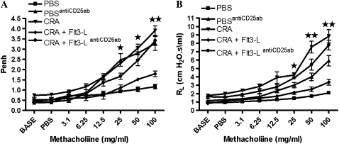
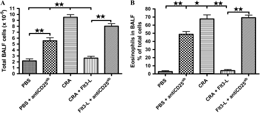
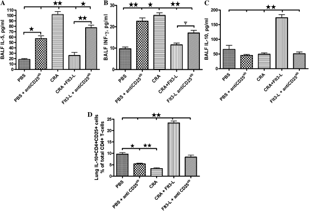
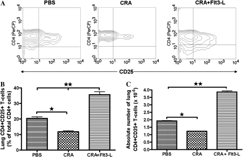
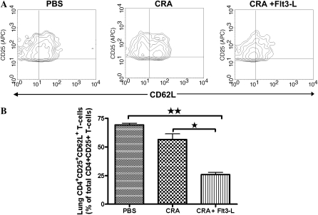
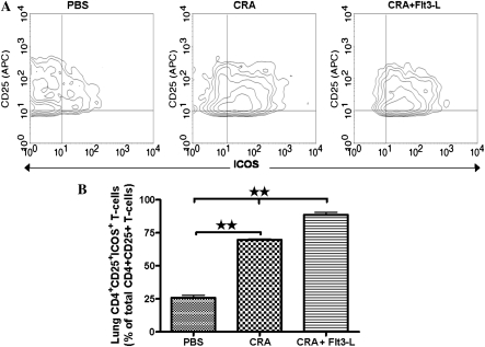
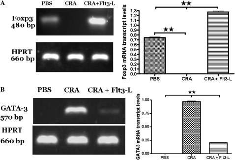
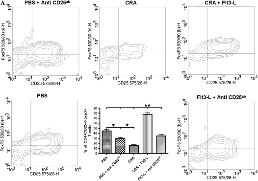
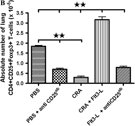
We previously reported in an ovalbumin-induced model of allergic asthma that Fms-like tyrosine kinase 3 ligand (Flt3-L) reversed airway hyperresponsiveness (AHR) and airway inflammation, and increased the number of regulatory CD11c(high)CD8 alpha(high)CD11b(low) dendritic cells in the lung. In this study, we investigated the effect of Flt3-L in a clinically relevant aeroallergen-induced asthma on the phenotypic expression of lung T cells. Balb/c mice were sensitized and challenged with cockroach antigen (CRA), and AHR to methacholine was established. These mice received three intraperitoneal injections of anti-CD25 antibody (PC61; 250 microg) and Flt3-L (3 microg) daily for 10 days. Cytokines and Ig levels in the serum were measured and differential bronchoalveolar lavage fluid (BALF) cell counts were examined. Flt3-L reversed AHR to methacholine to the control level. Flt3-L significantly decreased levels of BALF IL-5, IFN-gamma, eosinophilia and substantially increased IL-10 and the number of CD4(+)CD25(+) Forkhead winged helix transcription factor box P3 (Foxp3(+)) IL-10(+) T cells in the lung. Administration of PC61 antibody blocked the effect of Flt3-L and substantially increased AHR, eosinophilia, and BALF IL-5 and IFN-gamma levels, and decreased BALF IL-10 levels and the number of CD4(+)CD25(+)Foxp3(+)IL-10(+) T cells. Flt3-L significantly decreased CD62-L, but increased inducible costimulatory molecule and Foxp3 mRNA expression in the CD4(+)CD25(+) T cells isolated from lungs of Flt3-L-treated, CRA-sensitized mice compared to CRA-sensitized mice without Flt3-L treatment and PBS control group. Flt3-L significantly inhibited the effect of CRA sensitization and challenge to increase GATA3 expression in lung CD4(+)CD25(+) T cells. Collectively, these data suggest that the therapeutic effect of Flt3-L is mediated by increased density of naturally occurring CD4(+)CD25(+)Foxp3(+)IL-10(+)ICOS(+) T-regulatory cells in the lung. Flt3-L could be a therapeutic strategy for the management and prevention of allergic asthma.


Figures

Figure 1.
Figure 1.
Airway hyperresponsiveness (AHR) to methacholine. (A) On Day 33, AHR to methacholine (Mch) was established, followed by 10-day treatment with Fms-like tyrosine kinase 3 ligand (Flt3-L; 3 μg/d, intraperitoneal). On Day 44 AHR to Mch was measured, and enhanced pause (Penh) values were recorded. (B) On Day 45, airway resistance (RL) to Mch was recorded. Data are shown as mean (±SEM) for six mice in each group (*P < 0.01; **P < 0.001).
Figure 2.
Figure 2.
Bronchoalveolar lavage fluid (BALF) and cellularity. Recovered total cells in the BALF were counted, and differential analysis was performed using standard morphological criteria. A total of 300 cells were examined per cytospin slide, and absolute cell numbers were calculated per milliliter of the BALF based on the percentage of individual cells in a slide. Data are shown as mean (±SEM) for six animals in each group (**P < 0.001).
Figure 3.
Figure 3.
BALF cytokine level and IL-10–secreting CD4+CD25+ T-regulatory cells. BALF samples were immediately centrifuged and level of cytokines, IL-5 (A), IFNγ (B), and IL-10 (C), in the supernatants was measured. Data are shown as means (±SEM) for six animals in each group (*P < 0.01 and **P < 0.001). (D) CD4+CD25+ T cells were isolated, cultured, and plated on ELISPOT plates to evaluate expression of IL-10. Data are shown as means (±SEM) for five animals in each group (**P < 0.001).
Figure 4.
Figure 4.
Expression of CD4+CD25+ T cells. Contour plots show a comparative analysis of CD4+CD25+ expression of T cells isolated from lungs of PBS, cockroach antigen (CRA), and CRA/Flt3-L mice (A). Statistical analysis of the percentage of lung CD4+CD25+ T cells in each experimental group and Flt3-L–treated mice exhibits a significant increase of CD4+CD25+ T cell expression compared with CRA and PBS mice (B). Statistical analysis of the absolute number of lung CD4+CD25+ T cells in each experimental group is shown, and these data paralleled the percentage of CD4+CD25+ T cell (C) (*P < 0.01 and **P < 0.001).
Figure 5.
Figure 5.
Expression of CD62L on CD4+CD25+ T cells. Contour plots showed a comparative analysis of CD62L expression with CD4+CD25+ T cells isolated from lungs of PBS, CRA, and CRA/Flt3-L mice (A). Statistical analysis of CD62L expression of each experimental group and Flt3-L–treated mice exhibits a significant decrease of CD62L expression compared with CRA and PBS mice (B) (*P < 0.01 and **P < 0.001).
Figure 6.
Figure 6.
Expression of inducible costimulatory molecule (ICOS) on CD4+CD25+ T cells. Contour plots showed a comparative analysis of ICOS expression on CD4+CD25+ T cells isolated from lungs of PBS, CRA, and CRA/Flt3-L (A). Statistical analysis of ICOS expression of each experimental group and Flt3-L–treated mice exhibits a significant increase of ICOS expression compared with CRA and PBS mice (B) (**P < 0.001).
Figure 7.
Figure 7.
(A and B) The effect of Flt3-L on lung CD4+CD25+ T cells expressing Forkhead winged helix transcription factor box P3 (Foxp3) and GATA3. CD4+CD25+ T cell isolated from the lungs of PBS control mice showed normal expression of Foxp3 mRNA (480 bp) and no detectable level of GATA3 (570 bp). However, cockroach sensitization induced a significant decrease in Foxp3 expression, but substantially increased GATA3 expression in CD4+CD25+ T cell isolated from the lung tissue. Flt3-L caused a significant increase in Foxp3 and decreased expression of GATA3 compared with CRA mice and PBS control group. HPRT (660 bp) was used as the housekeeping gene. Densitometric analyses confirmed the PCR data by showing the ratio of mRNA intensity of Foxp3 and GATA3 by dividing by the intensity of the housekeeping gene, HPRT (**P < 0.001).
Figure 8.
Figure 8.
Intracellular protein expression of Foxp3 and the number of lung CD4+CD25+ after Flt3-L and PC61 treatment. (A) Contour plots of CD4+CD25+ T cells that are representative of all five groups. Foxp3 expression was significantly expressed in the PBS control compared with a substantial decrease in the CRA and PBS mice with PC61. Flt3-L–treated mice with PC61 showed significant decrease in Foxp3 on lung CD4+CD25+ T cells; however, Foxp3 was restored after Flt3-L treatment only (*P < 0.01; ΨP < 0.05; **P < 0.001). (B) In cockroach-sensitized and -challenged mice treated with Flt3-L, there was a significantly increased number of lung CD4+CD25+Foxp3+ T cells compared with sensitized mice without Flt3-L. This effect was completely inhibited by PC61, with a significant decrease in the number of lung CD4+CD25+Foxp3+ T cells (** P < 0.001).
Figure 8.
Figure 8.
Intracellular protein expression of Foxp3 and the number of lung CD4+CD25+ after Flt3-L and PC61 treatment. (A) Contour plots of CD4+CD25+ T cells that are representative of all five groups. Foxp3 expression was significantly expressed in the PBS control compared with a substantial decrease in the CRA and PBS mice with PC61. Flt3-L–treated mice with PC61 showed significant decrease in Foxp3 on lung CD4+CD25+ T cells; however, Foxp3 was restored after Flt3-L treatment only (*P < 0.01; ΨP < 0.05; **P < 0.001). (B) In cockroach-sensitized and -challenged mice treated with Flt3-L, there was a significantly increased number of lung CD4+CD25+Foxp3+ T cells compared with sensitized mice without Flt3-L. This effect was completely inhibited by PC61, with a significant decrease in the number of lung CD4+CD25+Foxp3+ T cells (** P < 0.001).

References

    1. Ohtomo T, Miyatake S, Kajiyama Y, Umezu-Goto M, Kobayashi N, Kaminuma O, Mori A. Airway eosinophilic inflammation is attenuated in conserved noncoding sequence-1–deficient mice. Int Arch Allergy Immunol 2008;146:2–6. - PubMed
    1. O'Sullivan SM. Asthma death, CD8+ T cells, and viruses. Proc Am Thorac Soc 2005;2:162–165. - PubMed
    1. Ramos-Barbon D, Presley JF, Hamid QA, Fixman ED, Martin JG. Antigen-specific CD4+ T cells drive airway smooth muscle remodeling in experimental asthma. J Clin Invest 2005;115:1580–1589. - PMC - PubMed
    1. Zhang-Hoover J, Finn P, Stein-Streilein J. Modulation of ovalbumin-induced airway inflammation and hyperreactivity by tolerogenic APC. J Immunol 2005;175:7117–7124. - PubMed
    1. Akbari O, Stock P, DeKruyff RH, Umetsu DT. Role of regulatory T cells in allergy and asthma. Curr Opin Immunol 2003;15:627–633. - PubMed

Publication types

MeSH terms