. 2009 Aug;182(4):1061-76.
doi: 10.1534/genetics.108.099986. Epub 2009 May 17.

A screen for modifiers of notch signaling uncovers Amun, a protein with a critical role in sensory organ development

Affiliations

A screen for modifiers of notch signaling uncovers Amun, a protein with a critical role in sensory organ development

Nevine A Shalaby et al. Genetics. 2009 Aug.

Abstract

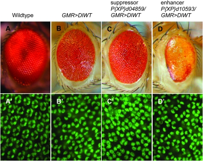
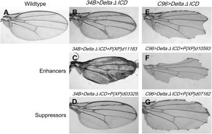
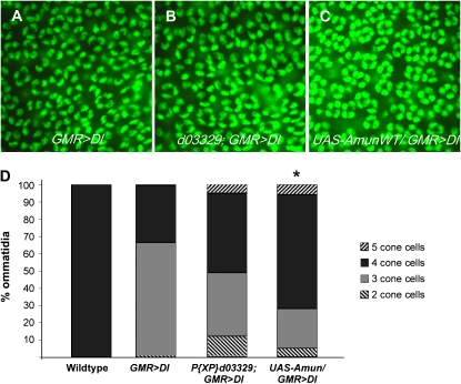
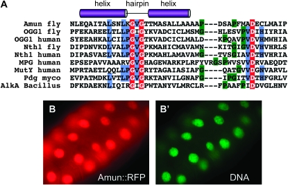
Notch signaling is an evolutionarily conserved pathway essential for many cell fate specification events during metazoan development. We conducted a large-scale transposon-based screen in the developing Drosophila eye to identify genes involved in Notch signaling. We screened 10,447 transposon lines from the Exelixis collection for modifiers of cell fate alterations caused by overexpression of the Notch ligand Delta and identified 170 distinct modifier lines that may affect up to 274 genes. These include genes known to function in Notch signaling, as well as a large group of characterized and uncharacterized genes that have not been implicated in Notch pathway function. We further analyze a gene that we have named Amun and show that it encodes a protein that localizes to the nucleus and contains a putative DNA glycosylase domain. Genetic and molecular analyses of Amun show that altered levels of Amun function interfere with cell fate specification during eye and sensory organ development. Overexpression of Amun decreases expression of the proneural transcription factor Achaete, and sensory organ loss caused by Amun overexpression can be rescued by coexpression of Achaete. Taken together, our data suggest that Amun acts as a transcriptional regulator that can affect cell fate specification by controlling Achaete levels.


Figures

F<sc>igure</sc> 1.—
Figure 1.—
Suppressors and enhancers of DeltaWT overexpression in the Drosophila eye. (A–D) Adult eyes. (A′–D′) Twenty-four-hour APF retinas stained with anti-Cut antibody to detect cone cells (green). (A and A′) A wild-type eye possesses an organized array of ommatidia (A); each ommatidium has four cone cells (A′). (B and B′) A GMR>DeltaWT/+ eye is small, glossy, and rough, with disorganized ommatidia (B) and an average of 3.34 cone cells/ommatidium (B′). (C and C′) A P{XP}d04859/GMR>DeltaWT eye. P{XP}d04859 mediates overexpression of Vha68-2. Adults have a larger, less glossy eye (C) with an average of 3.5 cone cells/ommatidium (C′); P < 0.005 compared with GMR>DlWT/+. (D and D′) A P{XP}d10593/GMR>DeltaWT eye. P{XP}d10593 mediates overexpression of Hr38. Adults have a smaller eye with loss of pigmentation and more disorganized ommatidia (D) with an average of 2.89 cone cells/ommatidium (D′); P < 0.005 compared with GMR>DlWT/+.
F<sc>igure</sc> 2.—
Figure 2.—
Secondary tests for confirmed modifiers of the GMR>DeltaWT/+ phenotype. (A) A wild-type adult wing. (B) Adult 34B-Gal4 UAS-DeltaΔICD/+ (34B>DeltaΔICD) wings display thickened wing veins, consistent with reduced Notch signaling. (C) Adult 34B>DeltaΔICD/+;P{XP}d11183/+ wings exhibit enhancement of the 34B>DeltaΔICD wing-vein-thickening phenotype. P{XP}d11183 disrupts karst. (D) Adult P{XP}d03329/+;34B>DeltaΔICD/+ wings exhibit suppression of the 34B>DeltaΔICD wing vein-thickening phenotype. P{XP}d03329 mediates overexpression of Amun and CG1837. (E) Adult UAS-DeltaΔICD/+;C96-Gal4/+ (C96>DeltaΔICD) wings display notches along the wing margin, typical of reduced Notch signaling. (F) Adult P{XP}d10593/UAS-DeltaΔICD;C96-Gal4/+ wings exhibit enhancement of the C96>DeltaΔICD wing notching phenotype. P{XP}d10593 mediates overexpression of Hr38. (G) Adult UAS-DeltaΔICD/+;C96-Gal4/P{XP}d07162 wings exhibit suppression of the C96>DeltaΔICD wing notching phenotype. P{XP}d07162 disrupts Cysteine string protein.
F<sc>igure</sc> 3.—
Figure 3.—
Overexpression of Amun suppresses the GMR>DeltaWT/+ cone cell phenotype. (A–C) Twenty-four-hour APF retinas stained with anti-Cut antibody (green) to detect cone cells. (A) A GMR>DeltaWT/+ retina exhibits a disorganized array of ommatidia with an average of 3.34 cone cells/ommatidium. (B) A P{XP}d03329/+;GMR>DeltaWT/+ retina exhibits suppression of the GMR>DeltaWT/+ cone cell phenotype, increasing the average to 3.5 cone cells/ommatidium. (C) A GMR>DeltaWT/+;UAS-AmunWT/+ retina exhibits greater suppression of the GMR>DeltaWT/+ cone cell phenotype, as compared to B, increasing the average to 3.73 cone cells/ommatidium (P < 0.005). (D) The proportional representation of ommatidia with two, three, four, or five cone cells. The percentage of ommatidia with four cone cells is greater for P{XP}d03329/+;GMR>DeltaWT/+ and GMR>DeltaWT/+;UAS-AmunWT/+ retinas than for GMR>DeltaWT/+ retinas. An asterisk denotes a statistically significant difference (P < 0.005) in average cone cells per ommatidium, as compared to GMR>DeltaWT/+.
F<sc>igure</sc> 4.—
Figure 4.—
Amun contains a putative DNA glycosylase domain and localizes to the nucleus. (A) Comparison of the Amun HhH DNA glycosylase domain to other known DNA glycosylases. The sequences used were the following: MPG, H. sapiens; MutY, H. sapiens; Pdg, M. tuberculosis; AlkA, B. subtilis; OGG1, D. melanogaster; Ogg1, H. sapiens; Nth1, D. melanogaster; Nth1, H. sapiens. There is little sequence similarity among these proteins; however, they share a conserved DNA-binding motif that consists of two α-helices (purple cylinders denote the approximate locations of these helices) connected by a hairpin loop with the consensus sequence LPG(V/I)G followed by a glycine/proline-rich region (green highlight) and a catalytically active aspartic acid residue (D, red highlight). The conserved H/N residue (blue highlight), following the catalytic D residue, differentiates between monofunctional (N) and bifunctional (H) glycosylases. Blue-highlighted L, P, and V residues are part of the consensus HhH domain. Red-highlighted white G residues are completely conserved in these DNA glycosylases. (B and B′) A dpp-Gal4/UAS-Amun∷RFP third larval instar salivary gland. Amun∷RFP (red) localizes to nuclei (B), as indicated by the DNA dye Vybrant Green (B′).
F<sc>igure</sc> 5.—
Figure 5.—
AmunRNAi effectively reduces Amun∷RFP protein expression. Third larval instar wing/notal imaginal discs. (A) A pnr-Gal4 UAS-Amun∷RFP/+ disc (pnr>Amun∷RFP). Overexpression of Amun∷RFP can be detected via RFP expression (red) in the pnr expression domain. (B) A UAS-AmunRNAi/+; pnr>Amun∷RFP/+ disc. AmunRNAi successfully reduces Amun∷RFP expression, as seen by the substantial reduction of Amun∷RFP signal within the pnr expression domain.
F<sc>igure</sc> 6.—
Figure 6.—
Amun loss-of-function, gain-of-function, and rescue experiments demonstrate a function for Amun during sensory organ development. (A) A wild-type adult notum. There are 10 organized rows of notal microchaetae including and between the two rows containing the dorsocentral macrochaetae (aDC and pDC). (B–D) Amun∷RFP overexpression phenotypes (assessed following growth at 27°). (E–H) Loss-of-function phenotypes that result from UAS-AmunRNAi expression (27° unless otherwise noted). (B) A ptc-Gal4/+;UAS-Amun∷RFP/+ notum results in loss of aSC (arrow) and pSC macrochaetae. (C) A sr-Gal4/UAS-Amun∷RFP notum exhibits severe loss of microchaetae in stripes 2 and 3 (highlighted with square brackets). (D) A pnr-Gal4 UAS-Amun∷RFP/+ notum exhibits severe loss of microchaetae across the central notum from stripe 1 to stripe 4 (highlighted with square brackets). (E) A UAS-AmunRNAi/+;UAS-Dicer2/ sca-Gal4 notum exhibits loss of aDC (data not shown), aSC (arrow), and pSC macrochaeta shafts (data not shown). (F) A UAS-AmunRNAi/+;UAS-Dicer2/ptc-Gal4 notum exhibits loss of aDC, pDC (arrow), and aSC (arrow) macrochaeta shafts, as well as misplacement of aSC macrochaetae. (G) A UAS-AmunRNAi/+;UAS-Dicer2/+;sr-Gal4/+ notum exhibits smaller microchaetae within the sr expression domain. Inset shows a magnified view of the medial notum. (H) A UAS-AmunRNAi/+;UAS-Dicer2/+;pnr-Gal4/+ notum (25°) displays smaller microchaetae and misplaced aDC macrochaetae. Inset shows a magnified view of the medial notum. (I) A UAS-AmunRNAi/+;pnr-Gal4 UAS-Amun∷RFP/+ notum. Co-expression of Amun∷RFP and AmunRNAi rescues the Amun∷RFP-induced gain-of-function phenotype (microchaeta loss) shown in D and the AmunRNAi-induced loss-of-function phenotype (smaller microchaetae) shown in H. (J) A UAS-Achaete/+;pnr-Gal4 UAS-Amun∷RFP/+ notum. Overexpression of Achaete partially rescues the Amun∷RFP overexpression phenotype (microchaeta loss) shown in D, supporting the hypothesis that loss of microchaetae occurs as a result of reductions in Achaete function. We also note the presence of ectopic macrochaetae, which may be due to Achaete overexpression. aDC, anterior dorsocentral; pDC, posterior dorsocentral; aSC, anterior scutellar; pSC, posterior scutellar.
F<sc>igure</sc> 7.—
Figure 7.—
Amun-induced loss of notal microchaetae is due to the loss of bristle organs and sensory organ precursors in the developing notum. (A and B) Nota from 31-hr APF pupae (27°) stained with MAb22C10 to detect neurons and shaft cells. (A) A wild-type notum has organized rows of microchaeta neurons and shaft cells. (B) A pnr-Gal4 UAS-Amun∷RFP/+ notum lacks staining for neurons within the pnr expression domain (outlined by dashes), indicating that the absence of external shafts is not due to the transformation of shaft/socket cells into neurons. (C and D) Nota from 15-hr APF pupae (27°) stained for β-galactosidase activity. The neur A101 lacZ insertion in the neuralized gene is used to mark SOPs. (C) A neur A101 notum exhibits wild-type rows of microchaete SOPs. (D) A pnr-Gal4 UAS-Amun∷RFP/neurA101 notum lacks SOPs within the pnr expression domain (outlined by dashes).
F<sc>igure</sc> 8.—
Figure 8.—
Amun overexpression downregulates Achaete levels in a cell-autonomous manner. (A and B) Nota from 9-hr APF pupae (27°) stained with anti-Achaete antibody (green). (A, A′, and A″) A sr-Gal/UAS-myr-mRFP notum. RFP (red) reflects expression of mRFP within the sr domain. Achaete-positive cells (arrows) mark the microchaeta proneural groups during this developmental stage. (B, B′, and B″) A sr-Gal4/UAS-Amun∷RFP notum shows a severe reduction or absence of Achaete expression (arrowheads) within the sr expression domain where Amun∷RFP is overexpressed. (C, C′, and C″) A 6- to 7-hr APF notum stained with anti-Achaete antibody (green in C and C″). Amun∷RFP is overexpressed randomly throughout the notal disc in Act5C-Gal4 clones (red). Achaete protein levels are downregulated in cells in which Amun∷RFP is overexpressed (arrowheads). Achaete-positive cells can be found directly adjacent to Amun∷RFP-positive cells (arrows) in clones in all regions of the notum.

Similar articles

Cited by

References

    1. Abdelilah-Seyfried, S., Y. M. Chan, C. Zeng, N. J. Justice, S. Younger-Shepherd et al., 2001. A gain-of-function screen for genes that affect the development of the Drosophila adult external sensory organ. Genetics 157 455–456. - PMC - PubMed
    1. Andersen, R., Y. Li, M. Resseguie and J. E. Brenman, 2005. Calcium/calmodulin-dependent protein kinase II alters structural plasticity and cytoskeletal dynamics in Drosophila. J. Neurosci. 25 8878–8888. - PMC - PubMed
    1. Artavanis-Tsakonas, S., 2004. Accessing the Exelixis collection. Nat. Genet. 36 207. - PubMed
    1. Artavanis-Tsakonas, S., M. D. Rand and R. J. Lake, 1999. Notch signalling: cell fate control and signal integration in development. Science 284 770–775. - PubMed
    1. Baker, K. D., L. M. Shewchuk, T. Kozlova, M. Makishima, A. Hassell et al., 2003. The Drosophila orphan nuclear receptor DHR38 mediates an atypical ecdysteroid signaling pathway. Cell 113 731–742. - PubMed

Publication types

MeSH terms