

The cyclin dependent kinase inhibitor (R)-roscovitine prevents alloreactive T cell clonal expansion and protects against acute GvHD
- PMID: 19448431
- PMCID: PMC2737335
- DOI: 10.4161/cc.8.11.8738
The cyclin dependent kinase inhibitor (R)-roscovitine prevents alloreactive T cell clonal expansion and protects against acute GvHD
Abstract
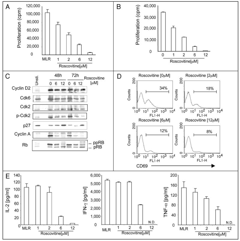
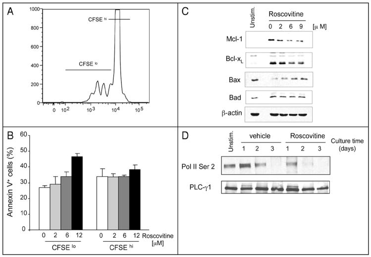
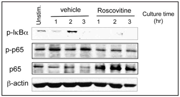
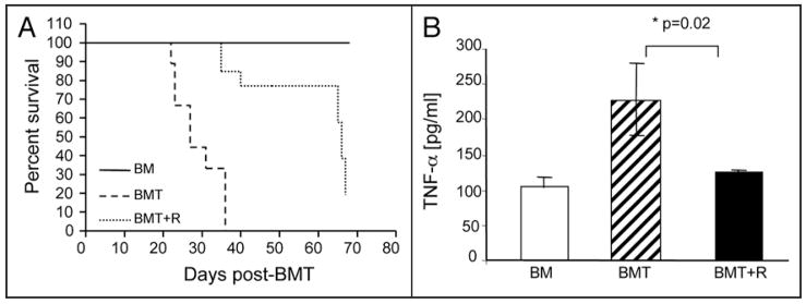
Cell cycle re-entry of quiescent T lymphocytes regulated by cdk2 is required for antigen-specific clonal expansion and generation of productive T cell responses. Recently, we determined that induction of antigen-specific T cell tolerance results in impaired cdk2 activity leading to enhanced Smad3 transactivation, upregulation of p15 and blockade of cell cycle progression. Here we report that pharmacologic inhibition of cdk2 with (R)-roscovitine blocked expansion of alloreactive T cells in vitro and in vivo and protected from lethal acute GvHD. In addition to inhibiting alloreactive T cell responses, (R)-roscovitine prevented TNF-alpha-mediated activation of NF-kappa B pathway, which is involved in the inflammatory process leading to the development of GvHD. The combined anti-proliferative and anti-inflammatory properties of (R)-roscovitine make it an attractive treatment modality toward control of GvHD.
Figures
Comment in
-
CDK inhibitors and immune responses.Cell Cycle. 2009 Aug 15;8(16):2484. doi: 10.4161/cc.8.16.9346. Cell Cycle. 2009. PMID: 19684461 No abstract available.
Similar articles
-
The cyclin dependent kinase inhibitor (R)-roscovitine mediates selective suppression of alloreactive human T cells but preserves pathogen-specific and leukemia-specific effectors.Clin Immunol. 2014 May-Jun;152(1-2):48-57. doi: 10.1016/j.clim.2014.02.015. Epub 2014 Mar 12. Clin Immunol. 2014. PMID: 24631965 Free PMC article.
-
Roscovitine suppresses CD4+ T cells and T cell-mediated experimental uveitis.PLoS One. 2013 Nov 18;8(11):e81154. doi: 10.1371/journal.pone.0081154. eCollection 2013. PLoS One. 2013. PMID: 24260551 Free PMC article.
-
CDK inhibitors and immune responses.Cell Cycle. 2009 Aug 15;8(16):2484. doi: 10.4161/cc.8.16.9346. Cell Cycle. 2009. PMID: 19684461 No abstract available.
-
Roscovitine confers tumor suppressive effect on therapy-resistant breast tumor cells.Breast Cancer Res. 2011 Aug 11;13(3):R80. doi: 10.1186/bcr2929. Breast Cancer Res. 2011. PMID: 21834972 Free PMC article.
-
Potential use of pharmacological cyclin-dependent kinase inhibitors as anti-HIV therapeutics.Curr Pharm Des. 2006;12(16):1949-61. doi: 10.2174/138161206777442083. Curr Pharm Des. 2006. PMID: 16787240 Review.
Cited by
-
The Role of Pharmacokinetics and Pharmacodynamics in Early Drug Development with reference to the Cyclin-dependent Kinase (Cdk) Inhibitor - Roscovitine.Sultan Qaboos Univ Med J. 2011 May;11(2):165-78. Epub 2011 May 15. Sultan Qaboos Univ Med J. 2011. PMID: 21969887 Free PMC article.
-
New roles for cyclin-dependent kinases in T cell biology: linking cell division and differentiation.Nat Rev Immunol. 2014 Apr;14(4):261-70. doi: 10.1038/nri3625. Epub 2014 Mar 7. Nat Rev Immunol. 2014. PMID: 24603166 Free PMC article. Review.
-
Cyclin-dependent kinase 5 activity is required for allogeneic T-cell responses after hematopoietic cell transplantation in mice.Blood. 2017 Jan 12;129(2):246-256. doi: 10.1182/blood-2016-05-702738. Epub 2016 Nov 14. Blood. 2017. PMID: 28064242 Free PMC article.
-
Acute Graft-versus-Host Disease - Biologic Process, Prevention, and Therapy.N Engl J Med. 2017 Nov 30;377(22):2167-2179. doi: 10.1056/NEJMra1609337. N Engl J Med. 2017. PMID: 29171820 Free PMC article. Review. No abstract available.
-
Foxp3 protein stability is regulated by cyclin-dependent kinase 2.J Biol Chem. 2013 Aug 23;288(34):24494-502. doi: 10.1074/jbc.M113.467704. Epub 2013 Jul 12. J Biol Chem. 2013. PMID: 23853094 Free PMC article.
References
-
- Appleman LJ, Boussiotis VA. T cell anergy and costimulation. Immunol Rev. 2003;192:161–80. - PubMed
-
- Williams MA, Bevan MJ. Effector and memory CTL differentiation. Annu Rev Immunol. 2007;25:171–92. - PubMed
-
- Sherr CJ, Roberts JM. Living with or without cyclins and cyclin-dependent kinases. Genes Dev. 2004;18:2699–711. - PubMed
-
- Matsuura I, Denissova NG, Wang G, He D, Long J, Liu F. Cyclin-dependent kinases regulate the antiproliferative function of Smads. Nature. 2004;430:226–31. - PubMed
MeSH terms
Substances
Grants and funding
LinkOut - more resources
Full Text Sources
Other Literature Sources