. 2009 Jul;44(7):375-83.
doi: 10.1097/RLI.0b013e3181a8afea.

High resolution ultra high field magnetic resonance imaging of glioma microvascularity and hypoxia using ultra-small particles of iron oxide

Affiliations

High resolution ultra high field magnetic resonance imaging of glioma microvascularity and hypoxia using ultra-small particles of iron oxide

Gregory A Christoforidis et al. Invest Radiol. 2009 Jul.

Abstract

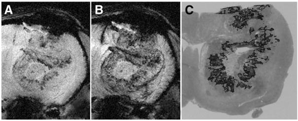
Objectives: This study assessed whether ultra-small particles of iron oxide (USPIO) intravascular contrast agent could enhance visualization of tumor microvascularity in F98 glioma bearing rats by means of ultra high field (UHF) high-resolution gradient echo (GRE) magnetic resonance imaging (MRI). In an effort to explain differences in visualization of microvascularity before and after USPIO administration, hypoxia and vessel diameters were assessed on corresponding histopathologic sections.

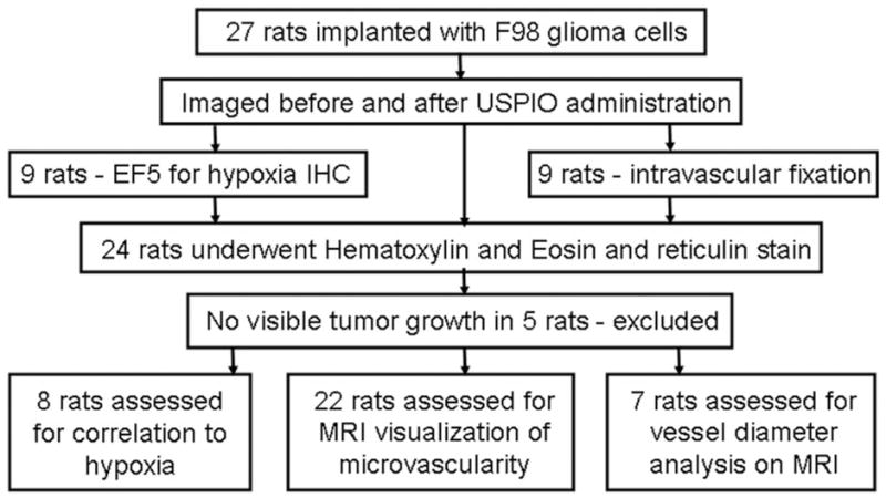
Materials and methods: F98 glioma cells were implanted stereotactically into the brains of syngeneic Fischer rats. Based on clinical criteria, rats were imaged 1 to 2 days before their death with and without USPIO contrast on an 8 Tesla MRI. To identify hypoxic regions of the brain tumor by immunohistochemical staining, a subset of animals also received a nitroimidazole-based hypoxia marker, EF5, before euthanasia. These sections then were compared with noncontrast enhanced MR images. The relative caliber of tumor microvasculature, compared with that of normal brain, was analyzed in a third group of animals.

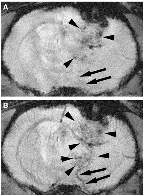
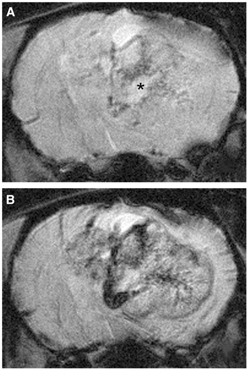
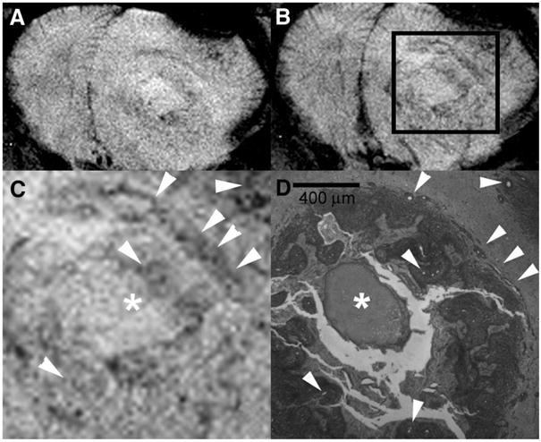
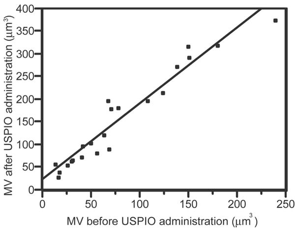
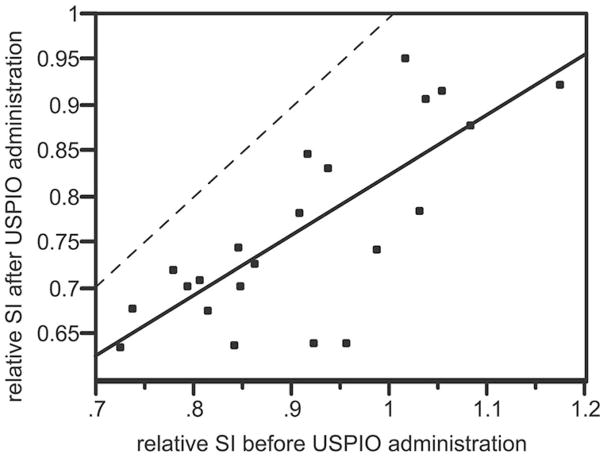
Results: After USPIO administration, UHF high-resolution GRE MRI consistently predicted increased microvascular density relative to normal gray matter when correlated with histopathology. The in-plane visibility of glioma microvascularity in 22 rats increased by an average of 115% and signal intensity within the tumor decreased by 13% relative to normal brain. Tumor microvascularity identified on noncontrast MR images matched hypoxic regions identified by immunohistochemical staining with a sensitivity of 83% and specificity of 89%. UHF GRE MRI was able to resolve microvessels less than 20 micro in diameter, although differences in tumor vessel size did not consistently account for differences in visualization of microvascularity.

Conclusions: USPIO administration significantly enhanced visualization of tumor microvascularity on gradient echo 8 T MRI and significantly improved visualization of tumor microvascularity. Microvascularity identified on precontrast images is suspected to be partly associated with hypoxia.


Figures

FIGURE 1
FIGURE 1
Overview of experiments. Rodents were implanted with F98 glioma cells and imaged. They were subsequently prepared for histopathology. A total of 20 rodents were included in the analysis (see text).
FIGURE 2
FIGURE 2
UHF GRE MRI of a tumor bearing rodent brain before (A) and after USPIO administration (B). Tumor microvessels are more readily identified on postcontrast images as serpiginous structures (arrowheads) that enhance after USPIO administration. The enlarged venous structure draining the tumor is more readily recognized on the postcontrast images (arrows). This draining vein is readily distinguished from tumor microvascularity (arrowheads). Note the relative enlargement of veins which drain tumor bed (arrows) compared with comparable vessels on the normal contralateral hemisphere.
FIGURE 3
FIGURE 3
Enhanced visualization of microvascularity within the F98 glioma using USPIO as an intravascular contrast agent. F98 glioma bearing rats were imaged without (A) and following USPIO administration (B). Note the enhanced visualization of hypointense serpiginous vessels within the tumor. Microvascularity within the noncontrast images surrounding the area of necrosis (asterisk, A) is where tumor hypoxia is typically found.
FIGURE 4
FIGURE 4
UHF GRE HR MRI of the brain of F98 glioma bearing rat before (A) and after (B) USPIO administration with magnification (C) of the area within the box (B) is compared with a corresponding histopathologic section (40×) using reticulin stain (D) in a rat that received an intracardiac injection of formalin. Arrowheads indicate vessels within the tumor and the asterisk depicts a focus of necrosis. Assessment of tumoral microvessel diameters measured on histopathology (D) indicate that there was often a difference in microvessel diameter between vessels identified on the precontrast images (A) and those identified only on the postcontrast images (B). Microvessels at least as small as 20 μm diameter are visualized.
FIGURE 5
FIGURE 5
UHF GRE HR MRI of a F98 glioma bearing rat before (A) and after (B) USPIO administration compared with EF5 IHC stain for hypoxia (red), overlaid on to corresponding H and E stained section (C). Note the concurrence between the regions of hypoxia identified with the EF5 stain (C) and the areas of microvascularity identified on the noncontrast 8 T image (A). Hypoxic regions primarily surround the central area of necrosis, Microvascularity along the periphery of the tumor identified on the postcontrast images does not correspond to the hypoxic regions.
FIGURE 6
FIGURE 6
Bivariate linear fit for calculated in-plane visibility of microvascularity (MV) before (x-axis) versus after (y-axis) USPIO administration. The derived linearly fitted equation is: post contrast MV = 1.67 (precontrast MV) + 28 (P < 0.0001; r-square = 0.93). This indicates that USPIO significantly increases visualization of microvessels using UHF HR GRE MRI.
FIGURE 7
FIGURE 7
Bivariate linear fit for ratio of signal intensity derived from entire volume of tumoral microvascularity relative to normal contralateral brain (rSI) before (x-axis) versus after (y-axis) USPIO administration. The derived linearly fitted equation is: post USPIO rSI = 0.67 (precontrast rSI) + 0.15 (P < 0.0001; r-square = 0.59). The dashed line represents the point at which the relative tumor signal loss before versus after USPIO administration would be equal. All points fall below the dashed line indicating that a greater concentration of USPIO is present within this tumor at the time of imaging. Because USPIO at the time of imaging is expected to be entirely intravascular, this suggests that tumor microvascular density within the F98 glioma is greater than that of normal tissue.

Similar articles

Cited by

References

    1. Burgess RE, Yu Y, Christoforidis GA, et al. Human leptomeningeal and cortical vascular anatomy of the cerebral cortex at 8 Tesla. J Comput Assist Tomogr. 1999;23:850–856. - PubMed
    1. Dashner RA, Kangarlu A, Clark DL, et al. Limits of 8-Tesla magnetic resonance imaging spatial resolution of the deoxygenated cerebral microvasculature. J Magn Reson Imaging. 2004;19:303–307. - PubMed
    1. Christoforidis GA, Bourekas EC, Baujan M, et al. High resolution MRI of the deep Brain vascular anatomy at 8 Tesla: susceptibility-based enhancement of the venous structures. J Comput Assist Tomogr. 1999;23:857–866. - PubMed
    1. Dashner RA, Chakeres DW, Kangarlu A, et al. MR Imaging visualization of the cerebral microvasculature: a comparison of live and postmortem studies at 8 T. AJNR. 2003;24:1881–1884. - PMC - PubMed
    1. Christoforidis GA, Kangarlu A, Abduljalil AM, et al. Susceptibility-based imaging of glioblastoma microvascularity at 8 T: correlation of MR imaging and postmortem pathology. AJNR. 2004;25:756–760. - PMC - PubMed

Publication types