. 2009 Jun 11;459(7248):837-41.
doi: 10.1038/nature08028. Epub 2009 May 17.

Two types of dopamine neuron distinctly convey positive and negative motivational signals

Affiliations

Two types of dopamine neuron distinctly convey positive and negative motivational signals

Masayuki Matsumoto et al. Nature. .

Abstract

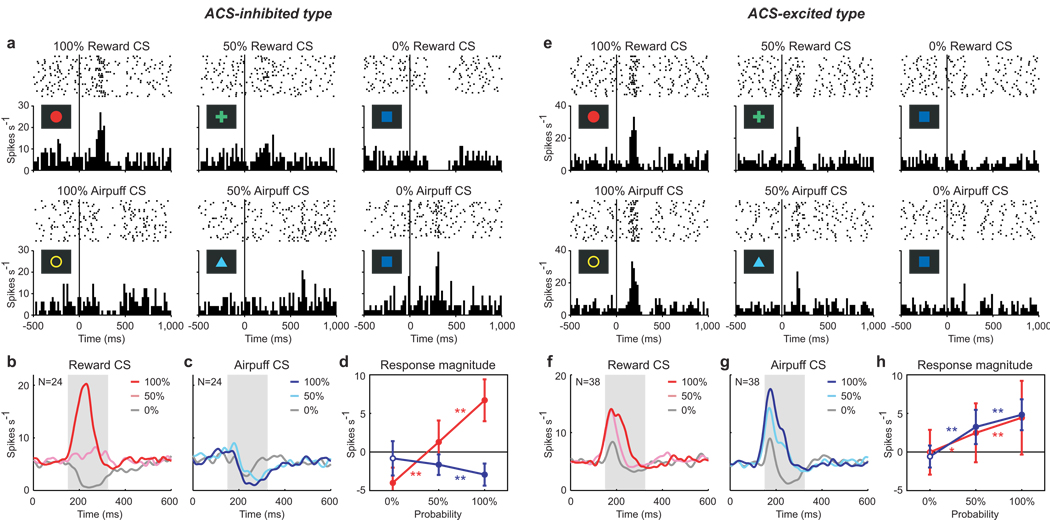
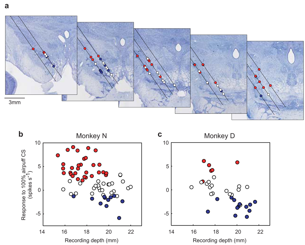
Midbrain dopamine neurons are activated by reward or sensory stimuli predicting reward. These excitatory responses increase as the reward value increases. This response property has led to a hypothesis that dopamine neurons encode value-related signals and are inhibited by aversive events. Here we show that this is true only for a subset of dopamine neurons. We recorded the activity of dopamine neurons in monkeys (Macaca mulatta) during a Pavlovian procedure with appetitive and aversive outcomes (liquid rewards and airpuffs directed at the face, respectively). We found that some dopamine neurons were excited by reward-predicting stimuli and inhibited by airpuff-predicting stimuli, as the value hypothesis predicts. However, a greater number of dopamine neurons were excited by both of these stimuli, inconsistent with the hypothesis. Some dopamine neurons were also excited by both rewards and airpuffs themselves, especially when they were unpredictable. Neurons excited by the airpuff-predicting stimuli were located more dorsolaterally in the substantia nigra pars compacta, whereas neurons inhibited by the stimuli were located more ventromedially, some in the ventral tegmental area. A similar anatomical difference was observed for their responses to actual airpuffs. These findings suggest that different groups of dopamine neurons convey motivational signals in distinct manners.


Figures

Figure 1
Figure 1. Pavlovian procedure
a, Appetitive block. Three conditioned stimuli (CSs) were associated with apple juice with 100%, 50% and 0% probability, respectively. b, Aversive block. Three CSs were associated with aversive airpuff with 100%, 50% and 0% probability, respectively. In both blocks, each trial started after the presentation of a timing cue (central small spot) on the screen. After 1 s, the timing cue disappeared and one of the three CSs was presented. After 1.5 s, the CS disappeared and the US (reward or airpuff) was delivered. In addition to the cued trials, uncued trials were included in which a reward alone (free reward) was delivered during the appetitive block and an airpuff alone (free airpuff) was delivered during the aversive block. c, Average of normalized magnitude of anticipatory licking during the presentation of reward CSs for monkey D (solid line) and monkey N (dashed line). d, Average of number of anticipatory blinks during the presentation of the airpuff CSs for monkey D (solid line) and monkey N (dashed line). Double asterisks indicate a significant difference between two data points (P < 0.01, Wilcoxon rank-sum test). Error bars indicate s.d.
Figure 2
Figure 2. Responses of dopamine neurons to CSs
a, e, Activity of two example neurons in the appetitive block (top row) and aversive block (bottom row) which were classified as ACS-inhibited type (a) and ACS-excited type (e). Histograms (20ms bins) and rasters are aligned at the onset of the CS and are shown for 100% reward CS, 50% reward CS, 0% reward CS, 100% airpuff CS, 50% airpuff CS, and 0% airpuff CS. b, c, Averaged activity of 24 ACS-inhibited type neurons. f, g, Averaged activity of 38 ACS-excited type neurons. Spike density functions (SDFs) are shown for 100% reward CS (dark red), 50% reward CS (light red) and 0% reward CS (gray) in the appetitive block (b, f), and for 100% airpuff CS (dark blue), 50% airpuff CS (light blue) and 0% airpuff CS (gray) in the aversive block (c, g). Gray area indicates the period that was used to analyze CS-evoked response. d, h, The magnitudes of the responses of the ACS-inhibited type neurons (d) and ACS-excited type neurons (h) to the reward CSs (red) and airpuff CSs (blue). Filled symbols indicate a significant deviation from zero (P < 0.05, Wilcoxon signed-rank test). Red and blue asterisks indicate a significant difference between two responses for reward and airpuff CSs, respectively (double asterisks, P < 0.01; single asterisks, P < 0.05, Wilcoxon signed-rank test). Error bars indicate s.d.
Figure 3
Figure 3. Responses of dopamine neurons to USs
a, e, Activity of two example neurons in the appetitive block (top row) and aversive block (bottom row) which were classified as AUS-inhibited type (a) and AUS-excited type (e). Histograms and rasters are aligned at the onset of the US and are shown for 100% reward, 50% reward, free reward, 100% airpuff, 50% airpuff, and free airpuff. b, c, Averaged activity of 47 AUS-inhibited type neurons. f, g, Averaged activity of 11 AUS-excited type neurons. SDFs are shown for 100% reward (dark red), 50% reward (light red) and free reward (gray) in the appetitive block (b, f), and for 100% airpuff (dark blue), 50% airpuff (light blue) and free airpuff (gray) in the aversive block (c, g). Gray area indicates the period that was used to analyze US-evoked response. d, h, The magnitudes of the responses of the AUS-inhibited type neurons (d) and AUS-excited type neurons (h) to reward (red) and airpuff (blue). Conventions are the same as Fig. 2d and h.
Figure 4
Figure 4. Locations of dopamine neurons in relation to their responses to airpuff-predicting CS
a, Recording sites of 68 dopamine neurons in monkey N are plotted on five coronal sections shown rostrocaudally from left to right (interval: 1 mm). Red circles indicate neurons showing significant excitations to 100% airpuff CS (i.e., ACS-excited type neurons). Blue circles indicate neurons showing significant inhibitions to 100% airpuff CS (i.e., ACS-inhibited type neurons). White circles, no significance (i.e., ACS-nonresponsive type neurons). Black lines indicate electrode penetration tracks which were tilted laterally by 35 degrees. b, c, Relation between recording depth and the response to 100% airpuff CS for monkey N (b) and monkey D (c). Red, blue, and white circles indicate ACS-excited, ACS-inhibited, and ACS-nonresponsive type neurons. The recording depth was measured from a reference depth set by a manipulator to advance the recording electrode.

References

    1. Schultz W. Predictive reward signal of dopamine neurons. J. Neurophysiol. 1998;80:1–27. - PubMed
    1. Satoh T, Nakai S, Sato T, Kimura M. Correlated coding of motivation and outcome of decision by dopamine neurons. J. Neurosci. 2003;23:9913–9923. - PMC - PubMed
    1. Takikawa Y, Kawagoe R, Hikosaka O. A possible role of midbrain dopamine neurons in short- and long-term adaptation of saccades to position-reward mapping. J. Neurophysiol. 2004;92:2520–2529. - PubMed
    1. Morris G, Arkadir D, Nevet A, Vaadia E, Bergman H. Coincident but distinct messages of midbrain dopamine and striatal tonically active neurons. Neuron. 2004;43:133–143. - PubMed
    1. Tobler PN, Fiorillo CD, Schultz W. Adaptive coding of reward value by dopamine neurons. Science. 2005;307:1642–1645. - PubMed

Publication types