

doi: 10.1038/nm.1959.
Netting neutrophils in autoimmune small-vessel vasculitis
Affiliations
- PMID: 19448636
- PMCID: PMC2760083
- DOI: 10.1038/nm.1959
Item in Clipboard
Netting neutrophils in autoimmune small-vessel vasculitis
Nat Med.
2009 Jun.
Abstract
Small-vessel vasculitis (SVV) is a chronic autoinflammatory condition linked to antineutrophil cytoplasm autoantibodies (ANCAs). Here we show that chromatin fibers, so-called neutrophil extracellular traps (NETs), are released by ANCA-stimulated neutrophils and contain the targeted autoantigens proteinase-3 (PR3) and myeloperoxidase (MPO). Deposition of NETs in inflamed kidneys and circulating MPO-DNA complexes suggest that NET formation triggers vasculitis and promotes the autoimmune response against neutrophil components in individuals with SVV.
Figures
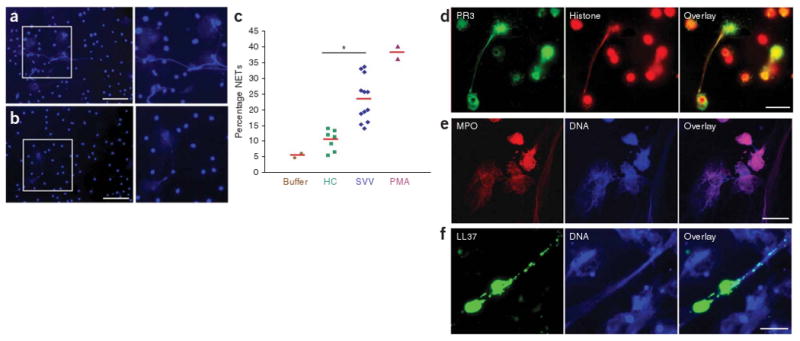
ANCA-induced formation of NETs containing autoantigens PR3 and MPO. (a,b) Fluorescence imaging of isolated tumor necrosis factor-α–primed neutrophils incubated with ANCA IgG (a) and control IgG (b) and stained for DNA. Scale bars, 50 μm. (c) Quantification of NET formation by fluorescence microscopy analysis. The percentage of cells with enlarged nuclei producing extracellular DNA fibers after 180 min incubation with buffer control, IgG from healthy controls (HC; n = 7), IgG from individuals with SVV (n = 12), or PMA as positive control is shown. Red horizontal bars indicate average percentage of each group. *P < 0.05. (d–f) Immunofluorescence analysis of autoantigens and immunostimulatory LL37 on PMA-induced NETs. (d) Immunofluorescence analysis of NETs stained with histone-specific antibody (red) and PR3-specific antibody (green). (e) Immunofluorescence analysis of NETs stained with MPO-specific antibody (red) and DNA stained with Hoechst dye (blue). (f) Immunofluorescence analysis of NET-forming neutrophils stained with LL37-specific antibody (green) and DNA stained with Hoechst dye (blue). Scale bars, 5 μm.
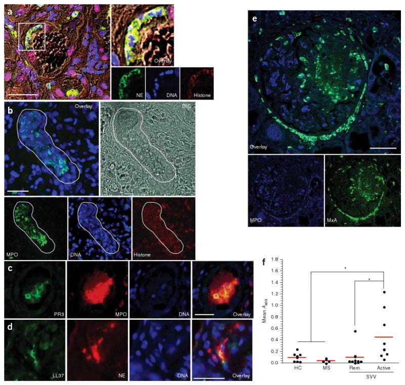
In vivo evidence for NET formation in individuals with SVV. (a–d) In situ immunofluorescence analysis of paraffin-embedded tissue sections of kidney needle biopsies from individuals with SVV glomerulonephritis. Tissue structure was visualized by differential interference contrast (DIC) microscopy. NETs were identified by colocalization of DNA, histone and neutrophil granule markers. (a) Colocalization of DNA (blue), histone (red) and neutrophil elastase (green) indicates intraglomerular NET formation. Blow up of boxed area shows NET deposition inside glomerular capsule. (b) Confocal immunofluorescence analysis showing large areas of colocalized DNA in blue, histone in red and MPO in green (defined by white line), indicating deposition of NETs that show autoantigen in the inflamed kidney of individuals with SVV. (c) Colocalization of MPO (red), PR3 (green) and DNA (blue), showing NET-autoantigen deposits inside the glomerulus. (d) Immunostaining of LL37 in kidney tissue of humans with SVV shows extracellularly located LL37 (green), DNA (blue) and NE (red), indicating NETs that are coated with LL37 and granular components. (e) MxA expression in close proximity to intra- and periglomerular neutrophil infiltrates. A glomerulus with extensive segmental necrosis and with pronounced extracellular MPO (green) deposition is shown. Numerous MxA (blue)-positive cells are scattered around MPO-positive neutrophil infiltrates and extracellular MPO deposits in the necrotic area. Tubular epithelia are partially MxA positive. Scale bars, 50 μm (a) and 25 μm (b-e). (f) Quantification of MPO-DNA complexes in the serum samples. The mean optical density as measured by capture ELISA using serum samples from healthy donors (n = 9), individuals with multiple sclerosis (MS; n = 5) and individuals with SVV in remission (Rem.; n = 10) and with active disease (n = 9) is shown; *P < 0.05. This study was approved by the local ethical committee of the Ludwig Maximilian University in Munich.
Comment in
-
LAMPs and NETs in the pathogenesis of ANCA vasculitis.J Am Soc Nephrol. 2009 Aug;20(8):1654-6. doi: 10.1681/ASN.2009060616. Epub 2009 Jul 16. J Am Soc Nephrol. 2009. PMID: 19608698 No abstract available.
References
Publication types
MeSH terms
Substances
Grants and funding
LinkOut - more resources
Full Text Sources
Other Literature Sources
Medical
Research Materials
Miscellaneous
