

c-Myc activates multiple metabolic networks to generate substrates for cell-cycle entry
- PMID: 19448666
- PMCID: PMC2779836
- DOI: 10.1038/onc.2009.112
c-Myc activates multiple metabolic networks to generate substrates for cell-cycle entry
Abstract
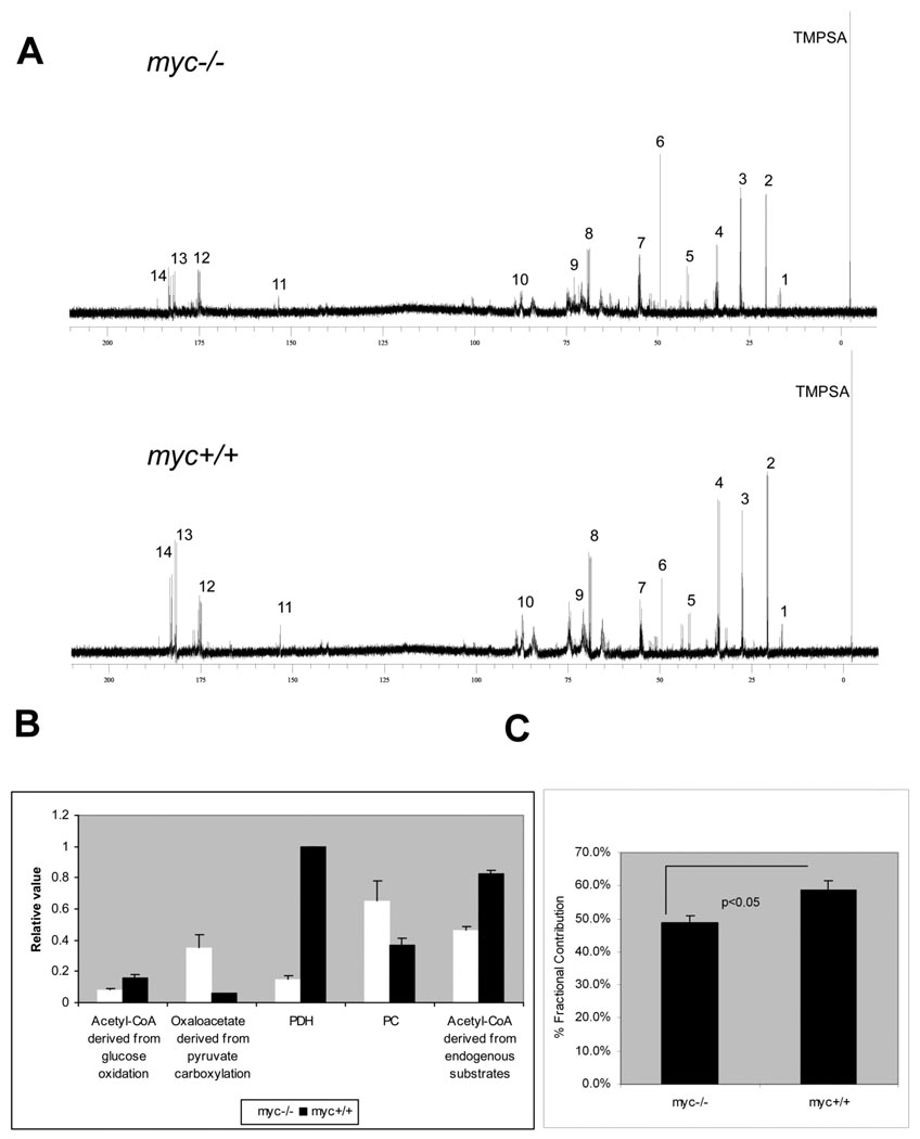
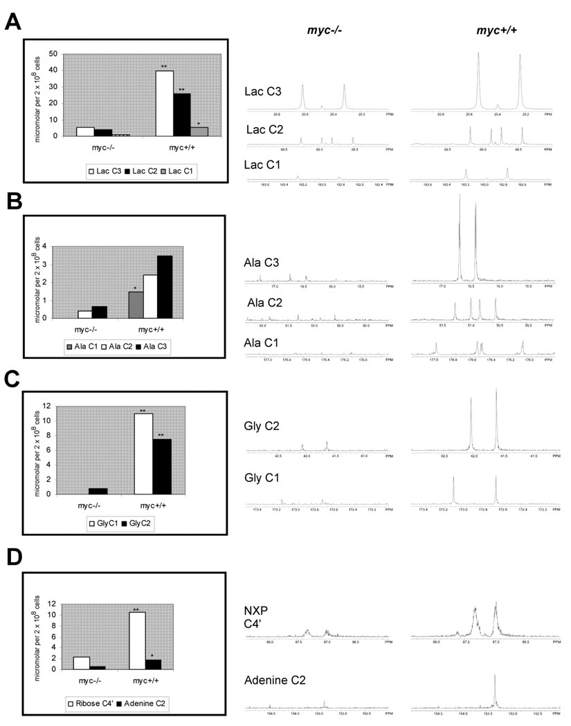
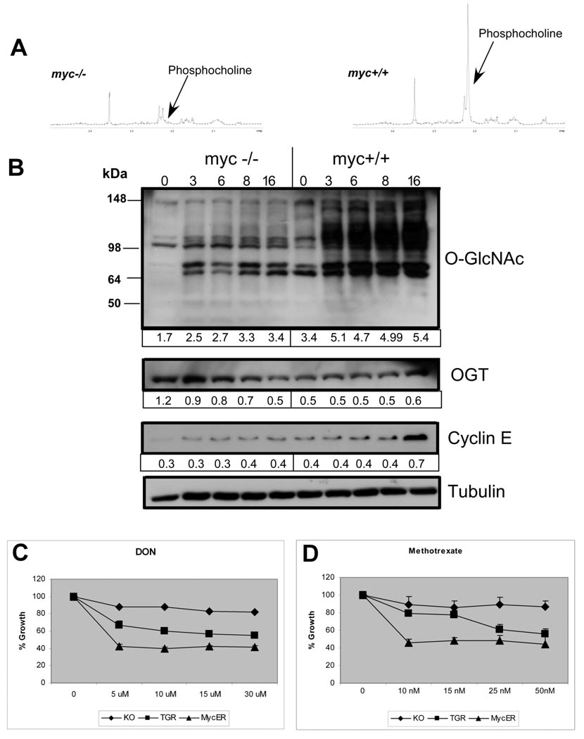
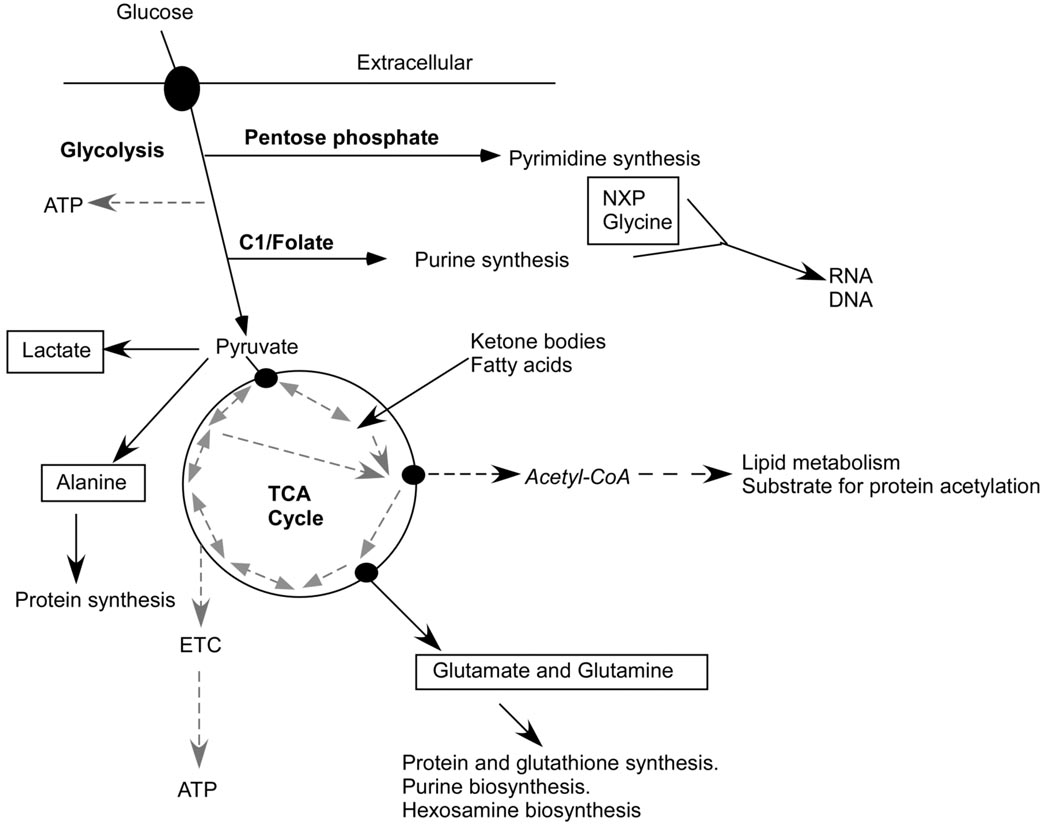
Cell proliferation requires the coordinated activity of cytosolic and mitochondrial metabolic pathways to provide ATP and building blocks for DNA, RNA and protein synthesis. Many metabolic pathway genes are targets of the c-myc oncogene and cell-cycle regulator. However, the contribution of c-Myc to the activation of cytosolic and mitochondrial metabolic networks during cell-cycle entry is unknown. Here, we report the metabolic fates of [U-(13)C] glucose in serum-stimulated myc(-/-) and myc(+/+) fibroblasts by (13)C isotopomer NMR analysis. We demonstrate that endogenous c-myc increased (13)C labeling of ribose sugars, purines and amino acids, indicating partitioning of glucose carbons into C1/folate and pentose phosphate pathways, and increased tricarboxylic acid cycle turnover at the expense of anaplerotic flux. Myc expression also increased global O-linked N-acetylglucosamine protein modification, and inhibition of hexosamine biosynthesis selectively reduced growth of Myc-expressing cells, suggesting its importance in Myc-induced proliferation. These data reveal a central organizing function for the Myc oncogene in the metabolism of cycling cells. The pervasive deregulation of this oncogene in human cancers may be explained by its function in directing metabolic networks required for cell proliferation.
Figures
References
-
- Dang CV, O'Donnell KA, Zeller KI, Nguyen T, Osthus RC, Li F. The c-Myc target gene network. Semin Cancer Biol. 2006;16:253–264. - PubMed
-
- Dean M, Levine RA, Ran W, Kindy MS, Sonenshein GE, Campisi J. Regulation of c-myc transcription and mRNA abundance by serum growth factors and cell contact. J Biol Chem. 1986;261:9161–9166. - PubMed
-
- Hart GW, Housley MP, Slawson C. Cycling of O-linked beta-N-acetylglucosamine on nucleocytoplasmic proteins. Nature. 2007;446:1017–1022. - PubMed
-
- Hunt AN, Postle AD. Phosphatidylcholine biosynthesis inside the nucleus: is it involved in regulating cell proliferation? Adv Enzyme Regul. 2004;44:173–186. - PubMed
-
- Jackowski S. Coordination of membrane phospholipid synthesis with the cell cycle. J Biol Chem. 1994;269:3858–3867. - PubMed
Publication types
MeSH terms
Substances
Grants and funding
LinkOut - more resources
Full Text Sources
Other Literature Sources
