. 2009 Jun;34(3):389-95.
doi: 10.1139/H09-044.

Sarcoplasmic reticulum-mitochondrial through-space coupling in skeletal muscle

Affiliations

Sarcoplasmic reticulum-mitochondrial through-space coupling in skeletal muscle

Robert T Dirksen. Appl Physiol Nutr Metab. 2009 Jun.

Abstract

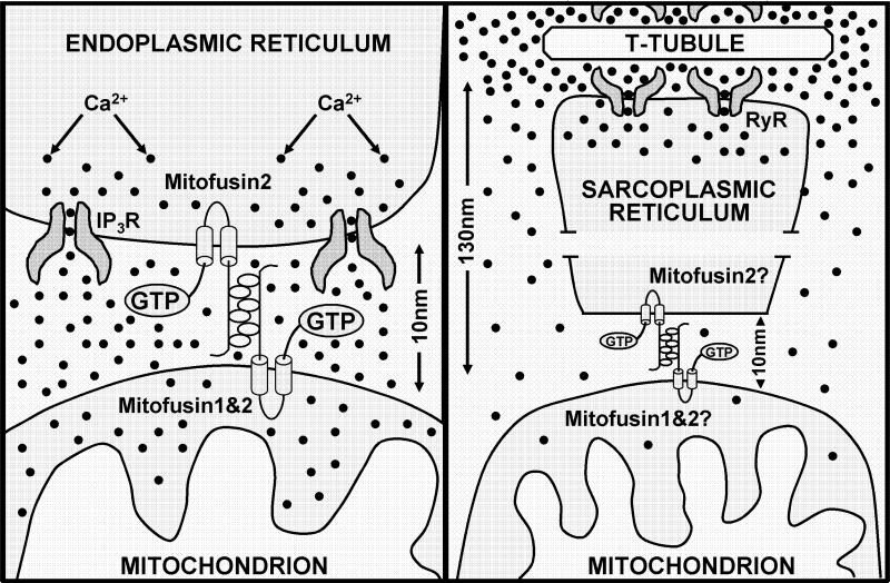
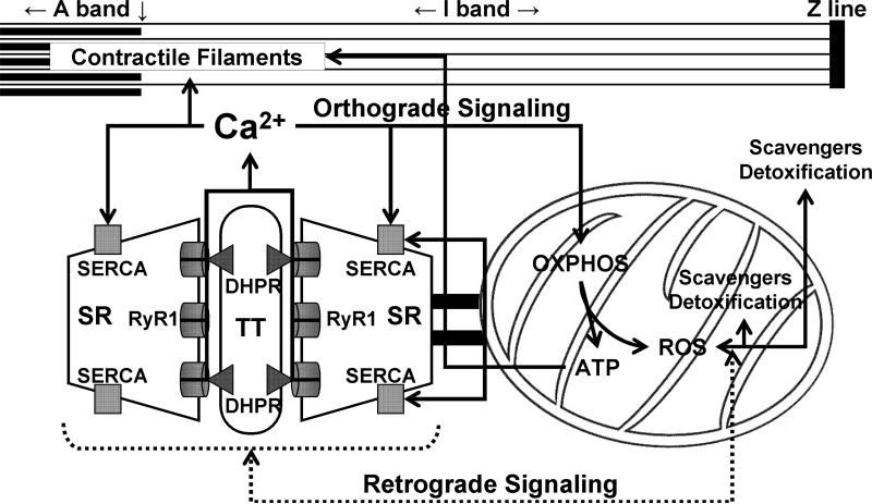
The skeletal muscle contractile machine is fueled by both calcium and ATP. Calcium ions activate the contractile machinery by binding to troponin C and relieving troponin-tropomyosin inhibition of actinomyosin interaction. ATP binding to myosin during the contractile cycle results in myosin detachment from actin, and energy liberated from subsequent ATP hydrolysis is then used to drive the next contractile cycle. ATP is also used to lower myoplasmic calcium levels during muscle relaxation. Thus, muscle contractility is intimately linked to the proper control of sarcomeric Ca2+ delivery and (or) removal and ATP generation and (or) utilization. In skeletal muscle, the sarcoplasmic reticulum (SR) is the primary regulator of calcium storage, release, and reuptake, while glycolysis and the mitochondria are responsible for cellular ATP production. However, the SR and mitochondrial function in muscle are not independent, as calcium uptake into the mitochondria increases ATP production by stimulating oxidative phosphorylation and mitochondrial ATP production, and production and (or) detoxification of reactive oxygen and nitrogen species (ROS/RNS), in turn, modulates SR calcium release and reuptake. Close spatial Ca2+/ATP/ROS/RNS communication between the SR and mitochondria is facilitated by the structural attachment of mitochondria to the calcium release unit (CRU) by 10 nm of electron-dense tethers. The resultant anchoring of mitochondria to the CRU provides a structural basis for maintaining bidirectional SR-mitochondrial through-space communication during vigorous contraction. This review will consider the degree to which this structural link enables privileged or microdomain communication between the SR and mitochondria in skeletal muscle.


Figures

Figure 1
Figure 1. Change in mitochondrial positioning during postnatal development
A-D) Representative electron micrographs depicting mitochondrial positioning in mouse FDB muscle fibers at 0.5 (A), 1 (B), 2 (C), and 4 (D) months after birth. Triads, small black arrows; mitochondria, open arrows; Z-line, large black arrows; SM, surface membrane. E and F) Representative high resolution electron micrographs showing mitochondrial association with the SR in mouse FDB muscle 1 (E) and 2 (F) months after birth. Small electron dense tethers (black arrows) bridge individual mitochondria (Mit.) to the SR. (G-I) Schematic representation of the parallel movement of the mitochondria and the CRU to the A-I band junction at 0.5 months after birth (G), 1 month after birth (H), and in adult skeletal muscle (I). Figure adapted with permission from Boncompagni, Rossi, Micaroni, Beznoussenko, Polishchuk, Dirksen, and Protasi, Mitochondria are Linked to Calcium Stores in Striated Muscle by Developmentally Regulated Tethering Structures, Molecular Biology Cell, 20(3):1058−1067, 2009.
Figure 2
Figure 2. Schematic representation of the dependence of microdomain Ca2+ signaling on mitochondrial location with respect to the site of Ca2+ release
(Left) Model of ER-mitochondrial localization and degree of microdomain Ca2+ signaling during IP3-mediated Ca2+ release (Merkwirth and Langer, 2008). When the mitochondrion is located only 10 nm from the point source of Ca2+ release, significant microdomain Ca2+ signaling occurs and mitochondrial Ca2+ uptake is driven by a large but rapidly dissipating local Ca2+ signal. Figure adapted from Cell, 135(7), Merkwirth and Langer, Mitofusin 2 builds a bridge between ER and mitochondria, 1165−1167, 2008, with permission from Elsevier. (Right) Model of SR-mitochondrial localization and degree of microdomain Ca2+ signaling during RyR1-mediated dependent Ca2+ release in adult mammalian skeletal muscle. Since CRU-associated mitochondria are located 130 nm from the point source of Ca2+ release (Boncompagni et al., 2009), limited microdomain signaling occurs and mitochondrial Ca2+ uptake is driven largely by the global myoplasmic Ca2+ signal.
Figure 3
Figure 3. Bidirectional SR-mitochondrial signaling in skeletal muscle
Ca2+ release during excitation-contraction (EC) coupling stimulates mitochondrial Ca2+ uptake and subsequent ATP production (Orthograde SR-mitochondrial signaling, solid lines). Mitochondrial-mediated Ca2+ spark suppression (Retrograde mitochondrial-SR signaling, broken lines) involves mitochondrial ROS scavenging and detoxification maintaining proper redox balance of the adjacent and tethered CRU. OXPHOS, oxidative phosphorylation; SR, sarcoplasmic reticulum; RyR1, type 1 ryanodine receptor; DHPR, dihydropyridine receptor; TT, transverse tubule; SERCA, sarco(endo)plasmic reticulum Ca2+-ATPase. Figure modified with permission from Rossi, Boncampagni, and Dirksen, Sarcoplasmic Reticulum-Mitochondrial Symbiosis: Bidirectional Signaling in Skeletal Muscle, Exercise and Sports Science Reviews, 37(1):29−35, 2009.

Similar articles

Cited by

References

    1. Allen DG, Lamb GD, Westerblad H. Skeletal muscle fatigue: cellular mechanisms. Physiol. Rev. 2008;88:287–332. - PubMed
    1. Balaban RS. Cardiac energy metabolism homeostasis: role of cytosolic calcium. J. Mol. Cell. Cardiol. 2002;34:1259–1271. - PubMed
    1. Beutner G, Sharma VK, Giovannucci DR, Yule DI, Sheu SS. Identification of a ryanodine receptor in rat heart mitochondria. J. Biol. Chem. 2001;276:21482–21488. - PubMed
    1. Boncompagni S, Rossi AE, Micaroni M, Beznoussenko GV, Polishchuk RS, Dirksen RT, Protasi F. Mitochondria are linked to calcium stores in striated muscle by developmentally regulated tethering structures. Mol Biol Cell. 2009;20:1058–1067. - PMC - PubMed
    1. Brookes PS, Yoon Y, Robotham JL, Anders MW, Sheu SS. Calcium, ATP, and ROS: a mitochondrial love-hate triangle. Am. J. Physiol. Cell Physiol. 2004;287:C817–C833. - PubMed

Publication types