Clinical Trial
. 2009 Dec;467(12):3307-20.
doi: 10.1007/s11999-009-0885-8. Epub 2009 May 16.

One-step bone marrow-derived cell transplantation in talar osteochondral lesions

Affiliations
Clinical Trial

One-step bone marrow-derived cell transplantation in talar osteochondral lesions

Sandro Giannini et al. Clin Orthop Relat Res. 2009 Dec.

Abstract

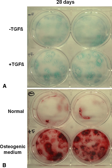
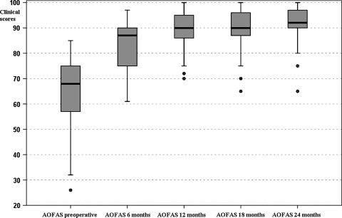
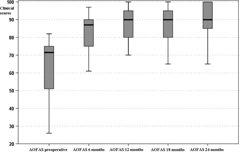
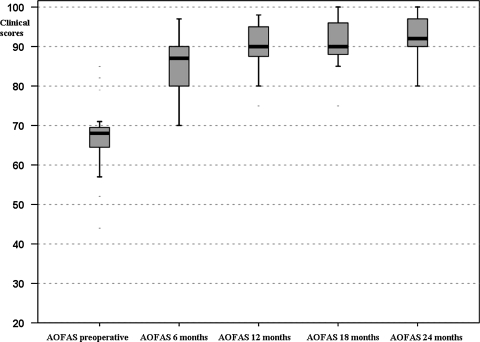
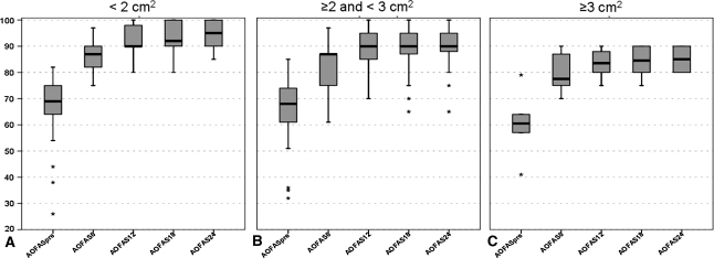
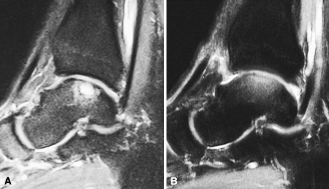
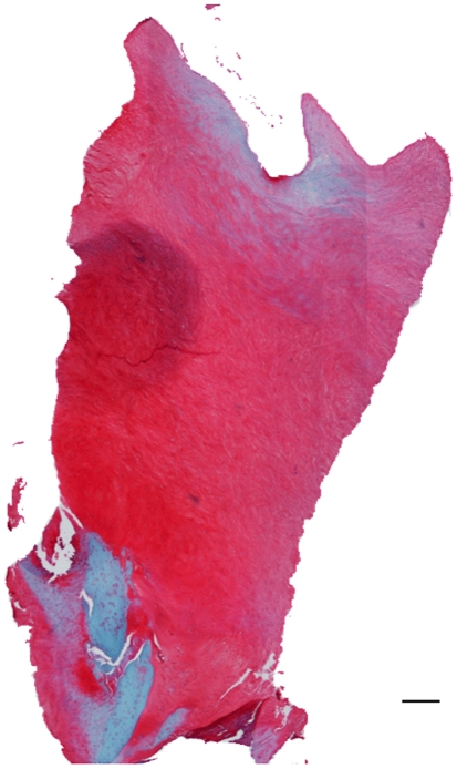
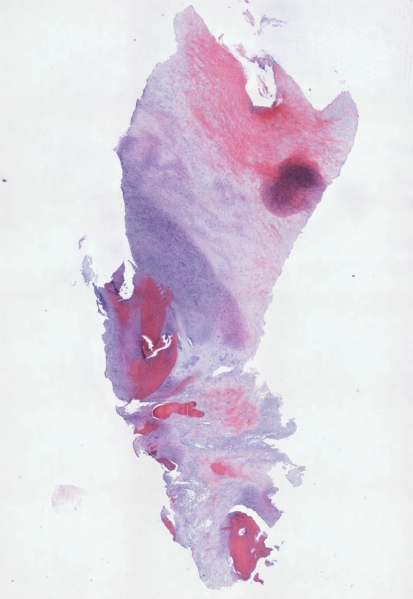
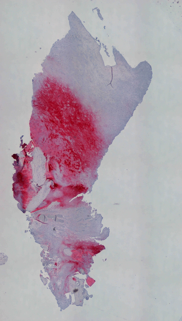
The ideal treatment of osteochondral lesions is debatable. Although autologous chondrocyte implantation provides pain relief, the need for two operations and high costs has prompted a search for alternatives. Bone marrow-derived cells may represent the future in osteochondral repair. Using a device to concentrate bone marrow-derived cells and collagen powder or hyaluronic acid membrane as scaffolds for cell support and platelet gel, a one-step arthroscopic technique was developed for cartilage repair. We performed an in vitro preclinical study to verify the capability of bone marrow-derived cells to differentiate into chondrogenic and osteogenic lineages and to be supported onto scaffolds. In a prospective clinical study, we investigated the ability of this technique to repair talar osteochondral lesions in 48 patients. Minimum followup was 24 months (mean, 29 months; range, 24-35 months). Clinical results were evaluated using the American Orthopaedic Foot and Ankle Society (AOFAS) score and the influence of scaffold type, lesion area, previous surgeries, and lesion depth was considered. MRI and histologic evaluation were performed. The AOFAS score improved from 64.4 +/- 14.5 to 91.4 +/- 7.7. Histologic evaluation showed regenerated tissue in various degrees of remodeling although none showed entirely hyaline cartilage. These data suggest the one-step technique is an alternative for cartilage repair, permitting improved functional scores and overcoming the drawbacks of previous techniques.

Level of evidence: Level IV, therapeutic study. See Guidelines for Authors for a complete description of levels of evidence.


Figures

Fig. 1A–D
Fig. 1A–D
The photographs show (A) the collagen powder seeded with concentrated cells, (B) staining of the collagen powder scaffold processed with MTT. The purple is indicative of the cells’ viability; (C) the hyaluronic acid membrane seeded with concentrated cells; and (D) staining of the hyaluronic acid membrane processed with MTT. The purple is indicative of the cells’ viability.
Fig. 2A–B
Fig. 2A–B
The photographs show (A) alcian blue staining of bone mineral-derived cells cultured in the absence (−) or presence (+) of TGF-β1 at 28 days and (B) alizarin red staining of bone mineral-derived cells cultured in normal (−) or osteogenic (+) medium at 28 days. Each evaluation was performed in duplicate.
Fig. 3
Fig. 3
Box-and-whisker plots show AOFAS clinical scores preoperatively and at the different followups. Horizontal line = median; box = first to third quartile; error bars = minimum to maximum; dots = outliers.
Fig. 4
Fig. 4
Clinical results were analyzed by dividing patients in two groups according to scaffold type used. The box-and-whisker plots show AOFAS clinical scores preoperatively and at the different followups in the group treated with collagen powder scaffolds. Horizontal line = median; box = first to third quartile; error bars = minimum to maximum; dots = outliers.
Fig. 5
Fig. 5
Clinical results were analyzed by dividing patients in two groups according to scaffold type used. Box-and-whisker plots show AOFAS clinical scores preoperatively and at the different followups in the group treated with hyaluronic acid membrane scaffolds. Horizontal line = median; box = first to third quartile; error bars = minimum to maximum; dots = outliers.
Fig. 6A–C
Fig. 6A–C
The box-and-whisker plots show the relationship between AOFAS clinical score preoperatively and at the different followups and the area of the lesion by dividing the patients into three groups according to the area of the lesion: (A) < 2 cm2, (B) ≤ 2 and < 3 cm2, and (C) ≥ 3 cm2. Considering 2 cm2 and 3 cm2 as cutoff values, the three groups had different improvement curves. Horizontal line = median; box = first to third quartile; error bars = minimum to maximum; dots = outliers.
Fig. 7A–B
Fig. 7A–B
(A) A preoperative MR image of a 32-year-old man shows an osteochondral lesion of the talus. (B) At the 2-year followup, the patient’s MR image shows restoration of the cartilage layer and subchondral bone.
Fig. 8
Fig. 8
Histologic observations of a biopsy specimen from a representative patient treated with bone marrow-derived cell transplantation supported on a collagen powder scaffold show repair tissue 12 months after surgery. Safranin O staining shows various degrees of tissue remodeling with many fibrocartilaginous features. Proteoglycans are represented, whereas collagen fibers are limited to a very narrow zone of the superficial layer (Original magnification, ×40).
Fig. 9
Fig. 9
Hematoxylin and eosin staining of a biopsy specimen shows tissue that is highly cellularized and the cells are homogeneously distributed inside the extracellular matrix (Original magnification, ×40).
Fig. 10
Fig. 10
Immunohistochemical evaluation of collagen Type II, using a new fuchsin stain, showed that the tissue is positive to this specific marker particularly in the medial and deep zone (Original magnification, ×40).

References

    1. {'text': '', 'ref_index': 1, 'ids': [{'type': 'DOI', 'value': '10.1002/(SICI)1097-4636(199811)42:2<172::AID-JBM2>3.0.CO;2-M', 'is_inner': False, 'url': 'https://doi.org/10.1002/(sici)1097-4636(199811)42:2<172::aid-jbm2>3.0.co;2-m'}, {'type': 'PubMed', 'value': '9773813', 'is_inner': True, 'url': 'https://pubmed.ncbi.nlm.nih.gov/9773813/'}]}
    2. Aigner J, Tegeler J, Hutzler P, Campoccia D, Pavesio A, Hammer C, Kastenbauer E, Naumann A. Cartilage tissue engineering with novel nonwoven structured biomaterial based on hyaluronic acid benzyl ester. J Biomed Mater Res. 1998;42:172–181. - PubMed
    1. {'text': '', 'ref_index': 1, 'ids': [{'type': 'DOI', 'value': '10.1136/ard.51.9.1056', 'is_inner': False, 'url': 'https://doi.org/10.1136/ard.51.9.1056'}, {'type': 'PMC', 'value': 'PMC1004837', 'is_inner': False, 'url': 'https://pmc.ncbi.nlm.nih.gov/articles/PMC1004837/'}, {'type': 'PubMed', 'value': '1417137', 'is_inner': True, 'url': 'https://pubmed.ncbi.nlm.nih.gov/1417137/'}]}
    2. Altman RD, Kates J, Chun LE, Dear DD, Eyre D. Preliminary observations of chondral abrasions in a canine model. Ann Rheum Dis. 1992;51:1056–1062. - PMC - PubMed
    1. {'text': '', 'ref_index': 1, 'ids': [{'type': 'PubMed', 'value': '2613129', 'is_inner': True, 'url': 'https://pubmed.ncbi.nlm.nih.gov/2613129/'}]}
    2. Angermann P, Jensen P. Osteochondritis dissecans of the talus: long-term results of surgical treatment. Foot Ankle. 1989;10:161–163. - PubMed
    1. {'text': '', 'ref_index': 1, 'ids': [{'type': 'DOI', 'value': '10.1302/0301-620X.87B5.15905', 'is_inner': False, 'url': 'https://doi.org/10.1302/0301-620x.87b5.15905'}, {'type': 'PubMed', 'value': '15855365', 'is_inner': True, 'url': 'https://pubmed.ncbi.nlm.nih.gov/15855365/'}]}
    2. Bartlett W, Skinner JA, Gooding CR, Carrington RW, Flanagan AM, Briggs TW, Bentley G. Autologous chondrocyte implantation versus matrix-induced autologous chondrocyte implantation for osteochondral defects of the knee: a prospective, randomised study. J Bone Joint Surg Br. 2005;87:640–645. - PubMed
    1. {'text': '', 'ref_index': 1, 'ids': [{'type': 'DOI', 'value': '10.2106/JBJS.E.00033', 'is_inner': False, 'url': 'https://doi.org/10.2106/jbjs.e.00033'}, {'type': 'PubMed', 'value': '16452741', 'is_inner': True, 'url': 'https://pubmed.ncbi.nlm.nih.gov/16452741/'}]}
    2. Baums MH, Heidrich G, Schultz W, Steckel H, Kahl E, Klinger HM. Autologous chondrocyte implantation of the ankle: a 2- to 5-year follow-up. J Bone Joint Surg Am. 2006;88:303–308. - PubMed

Publication types

MeSH terms