. 2009 Sep;238(9):2327-9.
doi: 10.1002/dvdy.21973.

Identification of genes expressed preferentially in the developing peripheral margin of the optic cup

Affiliations

Identification of genes expressed preferentially in the developing peripheral margin of the optic cup

Jeffrey M Trimarchi et al. Dev Dyn. 2009 Sep.

Abstract

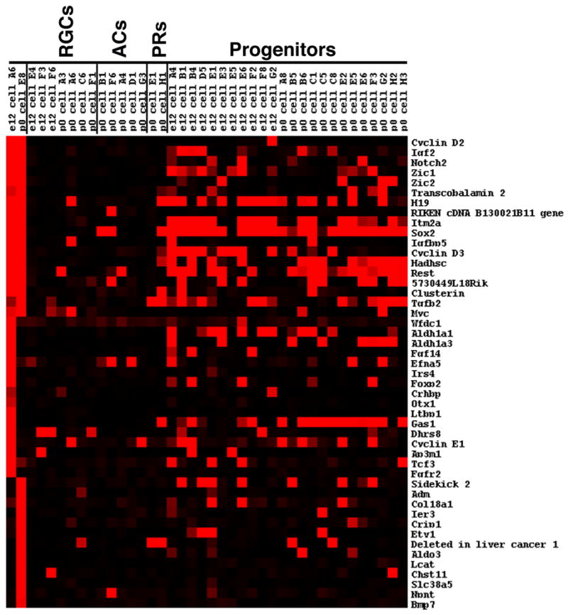
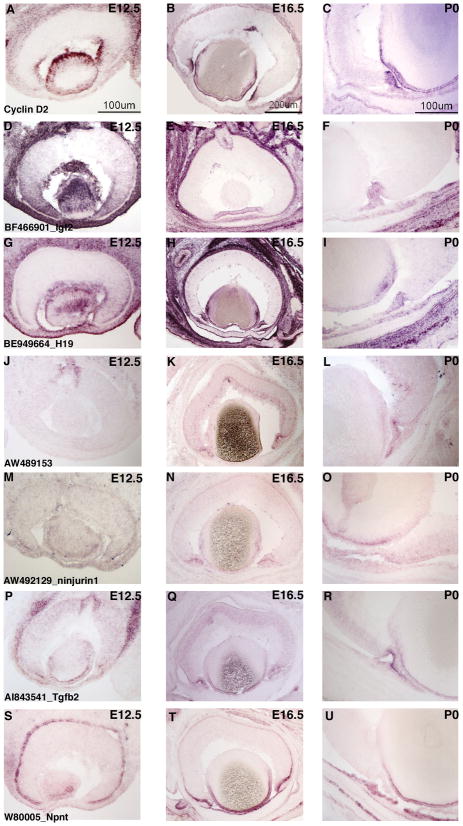
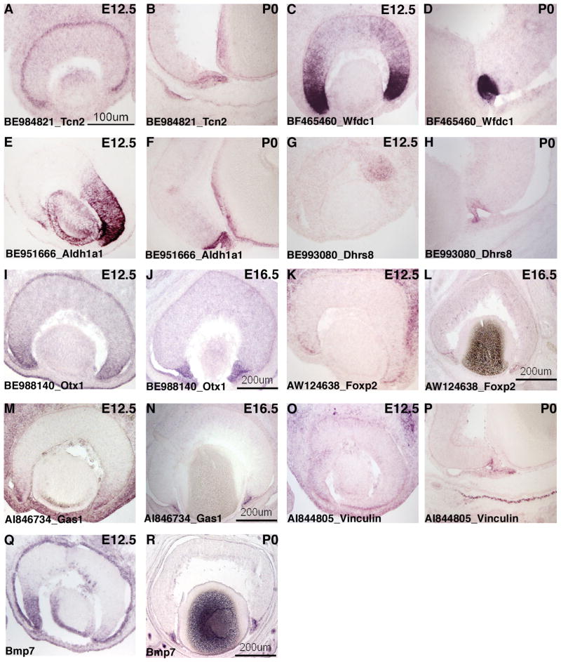
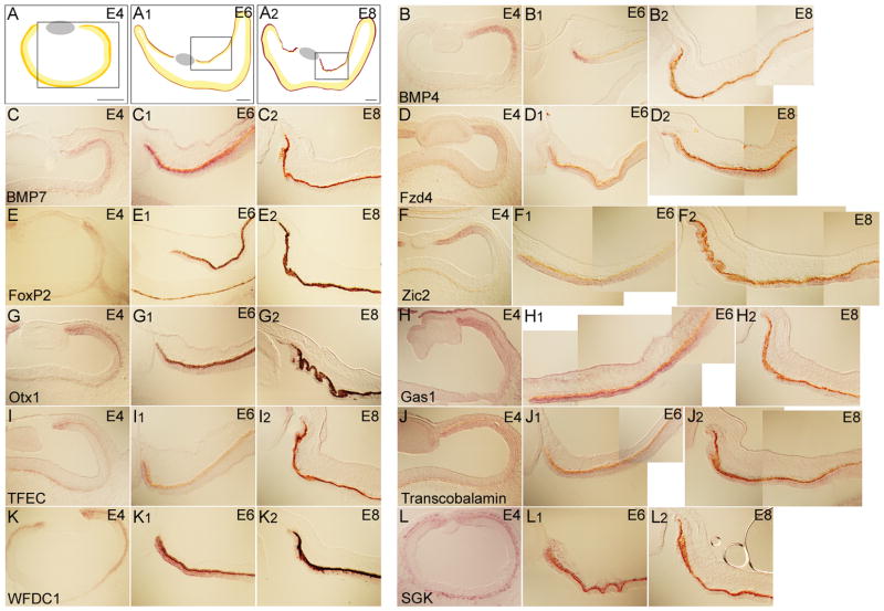
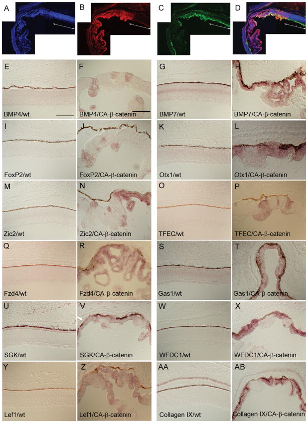
Specification of the peripheral optic cup by Wnt signaling is critical for formation of the ciliary body/iris. Identification of marker genes for this region during development provides a starting point for functional analyses. During transcriptional profiling of single cells from the developing eye, two cells were identified that expressed genes not found in most other single cell profiles. In situ hybridizations demonstrated that many of these genes were expressed in the peripheral optic cup in both early mouse and chicken development, and in the ciliary body/iris at subsequent developmental stages. These analyses indicate that the two cells probably originated from the developing ciliary body/iris. Changes in expression of these genes were assayed in embryonic chicken retinas when canonical Wnt signaling was ectopically activated by CA-beta-catenin. Twelve ciliary body/iris genes were identified as upregulated following induction, suggesting they are excellent candidates for downstream effectors of Wnt signaling in the optic cup.


Figures

Figure 1
Figure 1
Single cell transcriptional profiles of selected genes expressed in cells of the peripheral retina. The heatmap shown was generated using Treeview software and displays the expression of genes identified by visual inspection of the single cell microarray data. The expression is shown for the two new single cells that are putative ciliary body cells compared with individual developing ganglion cells (RGCs), developing amacrine cells (ACs), developing photoreceptors (PR), and cycling progenitor cells. The intensities of the Affymetrix signals have been scaled in the Treeview software such that signals of 10,000 and above are colored bright red, signals below 1,000 (designated as absent by the Affymetrix software) are black, and signals in between 10,000 and 1,000 are graded according to their signal value.
Figure 2
Figure 2
ISH analysis of candidate genes for the peripheral mouse eye. ISH on retinal cryosections was performed at three stages: E12.5 (A, D, G, J, M, P, S), E16.5 (B, E, H, K, N, Q, T) and P0 (C, F, I, L, O, R, U). The following probes were used: Cyclin D2 (AC), BF466901 [Igf2] (D–F), BE949664 [H19] (G–I), AW489153 (J–L), AW492129 [ninjurin1] (M–O) AI843541 [Tgfb2] (P–R), and W8005 [Npnt] (S–U). Representative scale bars are shown.
Figure 3
Figure 3
ISH analysis of candidate genes for the peripheral mouse eye. ISH on retinal cryosections was performed at: E12.5 (A, C, E, G, I, K, M, O, Q), E16.5 (J, L, N) and P0 (B, D, F, H, P, R). The following probes were used: BE984821 [Tcn2] (A,B), BF465460 [Wfdc1] (C,D), BE951666 [Aldh1a1] (E,F), BE993080 [Dhrs8] (G,H), BE988140 [Otx1] (I,J), AW124638 [Foxp2] (K,L), AI846734 [Gas1] (M,N), AI844805 [Vinculin] (O,P) and Bmp7 (Q,R). Representative scale bars are shown.
Figure 4
Figure 4
ISH analysis for the expression of eleven candidate genes for the peripheral chick eye. The scheme in A, A1 and A2 shows the area of peripheral eye represented in this figure for chick E4, E6 and E8, respectively. The probes used were BMP4 (B-B2), BMP7 (C-C2), Fzd4 (D-D2), FoxP2 (E-E2), Zic2 (F-F2), Otx1 (G-G2), Gas1 (H-H2), TFEC (I-I2), Transcobalamin 2 (J-J2), WFDC1 (K-K2), and SGK (L-L2). Scale bars: 150μm.
Figure 5
Figure 5
ISH analysis for the induction of peripheral marker genes upon canonical Wnt signaling activation. Optic vesicles were infected with a RCAS:CA-β-catenin at HH stage 10 and the analysis was performed on embryos harvested at E9.5. Using a representative section from the central part of the retina, the overlap between the virally infected region (B) and the area showing retinal thinning and folding [shown as the absence of β-tubulin antibody staining in (C)] is visualized in merged image (D). A DAPI-stained retinal section is shown in (A). The white arrowed line demarcates the normal uninfected portion of the retina. Probes used were BMP4 (E and F), BMP7 (G and H), FoxP2 (I and J), Otx1 (K and L), Zic2 (M and N), TFEC (O and P), Fzd4 (Q and R), Gas1 (S and T), SGK (U and V), WFDC1 (W and X), Lef1 (Y and Z), and Collagen IX (AA and AB). Images shown on the left are from the central retinal of uninfected chicks. The images on the right are also from the central retina of chicks infected with the RCAS:CA-β-catenin. The ventral side is down. Scale bars: 150μm.

Similar articles

Cited by

References

    1. Acampora D, Mazan S, Avantaggiato V, Barone P, Tuorto F, Lallemand Y, Brulet P, Simeone A. Epilepsy and brain abnormalities in mice lacking the Otx1 gene. Nat Genet. 1996;14:218–222. - PubMed
    1. Ahmad I, Dooley CM, Thoreson WB, Rogers JA, Afiat S. In vitro analysis of a mammalian retinal progenitor that gives rise to neurons and glia. Brain Res. 1999;831:1–10. - PubMed
    1. Araki T, Milbrandt J. Ninjurin, a novel adhesion molecule, is induced by nerve injury and promotes axonal growth. Neuron. 1996;17:353–361. - PubMed
    1. Beebe DC. Development of the ciliary body: a brief review. Trans Ophthalmol Soc U K. 1986;105 (Pt 2):123–130. - PubMed
    1. Blackshaw S, Harpavat S, Trimarchi J, Cai L, Huang H, Kuo WP, Weber G, Lee K, Fraioli RE, Cho SH, Yung R, Asch E, Ohno-Machado L, Wong WH, Cepko CL. Genomic analysis of mouse retinal development. PLoS Biol. 2004;2:E247. - PMC - PubMed

Publication types