. 2009 Jun;39(6):1619-31.
doi: 10.1002/eji.200839062.

A new function for LAT and CD8 during CD8-mediated apoptosis that is independent of TCR signal transduction

Affiliations

A new function for LAT and CD8 during CD8-mediated apoptosis that is independent of TCR signal transduction

Raedun L Clarke et al. Eur J Immunol. 2009 Jun.

Abstract

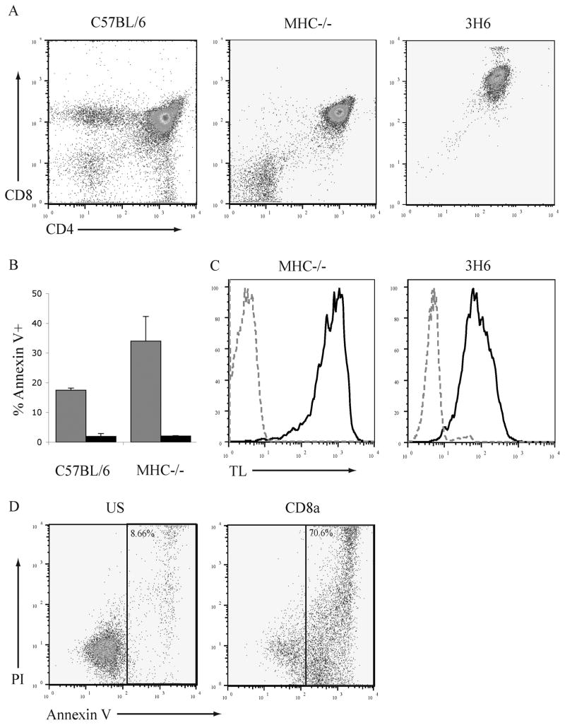
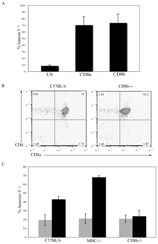
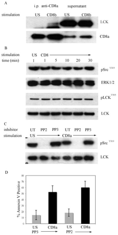
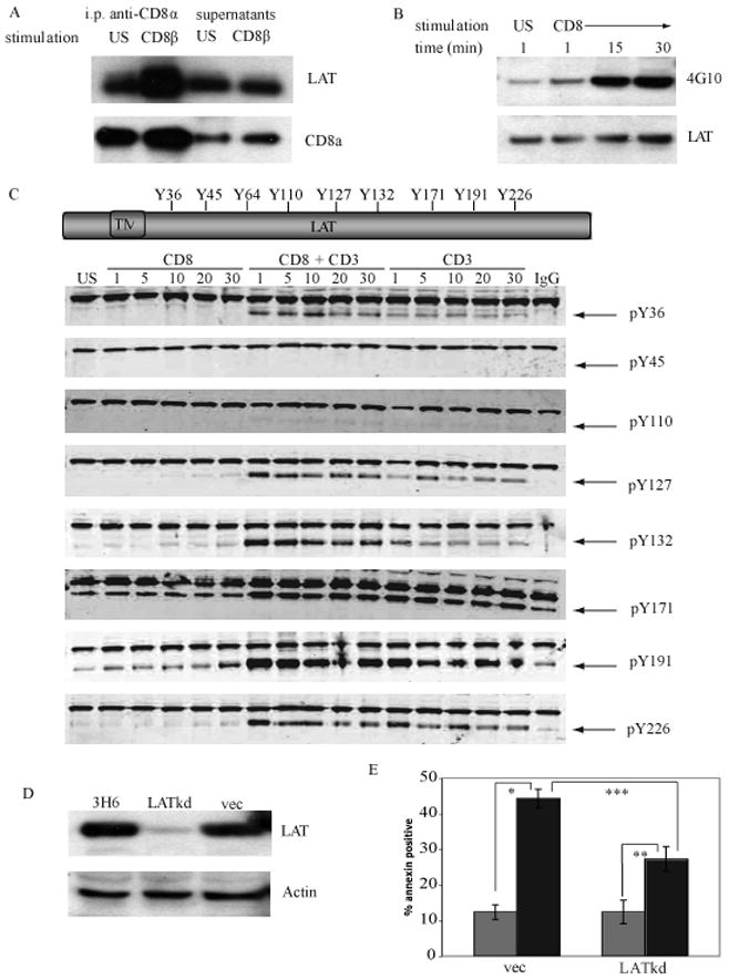
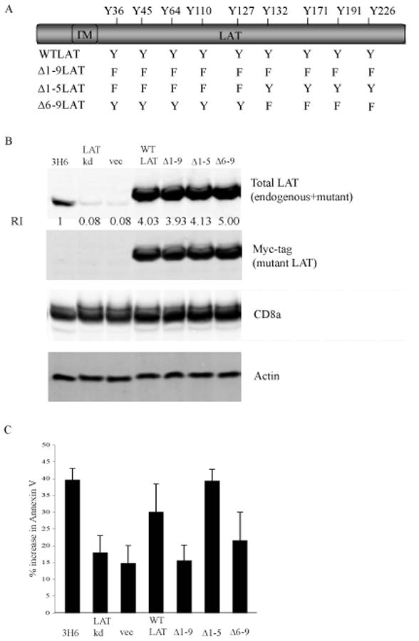
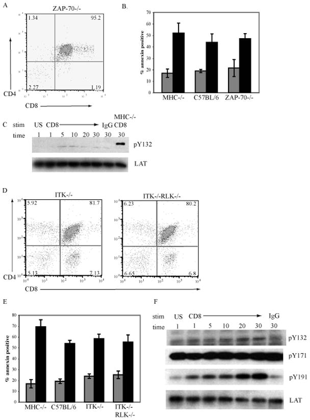
The majority (>95%) of thymocytes undergo apoptosis during selection in the thymus. Several mechanisms have been proposed to explain how apoptosis of thymocytes that are not positively selected occurs; however, it is unknown whether thymocytes die purely by "neglect" or whether signaling through a cell-surface receptor initiates an apoptotic pathway. We have previously demonstrated that on double positive thymocytes the ligation of CD8 in the absence of TCR engagement results in apoptosis and have postulated this is a mechanism to remove thymocytes that have failed positive selection. On mature single positive T cells CD8 acts as a co-receptor to augment signaling through the TCR that is dependent on the phosphorylation of the adaptor protein, linker for activation of T cells (LAT). Here, we show that during CD8-mediated apoptosis of double positive thymocytes there is an increase in the association of CD8 with LAT and an increase in LAT tyrosine phosphorylation. Decreasing LAT expression and mutation of tyrosine residues of LAT reduced apoptosis upon crosslinking of CD8. Our results identify novel functions for both CD8 and LAT that are independent of TCR signal transduction and suggest a mechanism for signal transduction leading to apoptosis upon CD8 crosslinking.


Conflict of interest statement

Conflict of Interest

The authors declare no commercial or financial conflict of interest.

Figures

Fig. 1
Fig. 1. The 3H6 thymoma cell line is susceptible to apoptosis induced by the ligation of CD8
A. Thymocytes from C57BL/6, MHC−/− mice, and 3H6 thymoma cells were stained with antibodies to CD4 and CD8α and analyzed by flow cytometry. B. Thymocytes from C57BL/6 and MHC−/− mice were treated with antibody to CD8α and goat anti-rat Ig (grey bars), CD4 and goat anti-rat Ig (hatched bars), or goat anti-rat Ig alone (black bars) for 6hr. Cells were stained with FITC-conjugated annexin V and analyzed by flow cytometry. N=3; mean ± SEM. C. Thymocytes from MHC−/− mice and 3H6 thymoma cells were stained with anti-CD4 (FITC), anti-CD8α (APC), and tetrameric TL molecules (PE) The binding of TL to DP cells is represented by the black line; unstained DP cells by the grey dotted line. D. 3H6 thymoma cells were treated with antibody to CD8α and goat anti-rat Ig, CD4 and goat anti-rat Ig, or left unstimulated (US) for 6hr. Cells were stained with annexin V (FITC) and PI, the percentage of the cells in the indicated gate is given.
Figure 2
Figure 2. The CD8β chain is required for CD8-mediated apoptosis
A. 3H6 thymoma cells were left unstimulated (US) or treated with anti-CD8α or anti-CD8β antibodies and goat anti-rat Ig to crosslink for 6 hours. Apoptosis was measured by the binding of annexin-V conjugated with FITC. N=4; mean ± SEM. B. Thymocytes from C57BL/6 and CD8β−/− mice were stained with anti-CD4 and anti-CD8 antibodies and analyzed by flow cytometry. C. Thymocytes from C57BL/6, MHC−/− and CD8β−/− mice were left US (grey bars) or crosslinked with anti-CD8α and goat anti-rat Ig (black bars) for 6 hours. Apoptosis was measured by the binding of annexin-V conjugated with FITC. N=5; mean ± SEM.
Figure 3
Figure 3. Lck is not required for CD8-mediated apoptosis of DP thymocytes
A. MHC−/− thymocytes were treated with antibody to CD8β and crosslinked with goat anti-rat Ig or left unstimulated (US) at 37°C for 30min. CD8α was immunoprecipitated and western blots of the immunoprecipitates and supernatants were probed with antibody to Lck and then reprobed with antibody to the intracellular tail of CD8α as a loading control. B. MHC−/− thymocytes were treated with antibody to CD8α and crosslinked with goat anti-rat Ig for the indicated times or left unstimulated (US). Western blots of whole cell lysates were analyzed for expression of pSrcY416 or pLckY505 and reprobed with antibodies to either ERK1/2 or Lck to monitor equivalent loading in each lane. C. MHC−/− thymocytes were treated with 5mM PP2, 5mM PP3, or left untreated (UT) for 30 minutes at 37°C. The thymocytes were then treated with anti-CD8α antibody and crosslinked with anti-rat Ig for 30 min at 37°C or left unstimulated. Western blots of whole cell lysates were analyzed with pSrcY416 antibody and reprobed with an anti-Lck antibody as a loading control. D. MHC−/− thymocytes that were treated with PP2 or PP3 were cross-linked with antibody to CD8α (black bars) or left unstimulated (grey bars). At 6 hours apoptosis was measured by the binding annexin-V conjugated with FITC. N=3; mean ± SEM.
Fig. 4
Fig. 4. LAT is required for CD8-mediated apoptosis of thymocytes
A. MHC−/− thymocytes were left unstimulated (US) or treated with antibody to CD8β and goat anti-rat Ig for 30 min. CD8 was immunoprecipitated with biotinylated antibody to CD8α coupled to Neutravidin beads for 90 min. Western blots of the immunoprecipitates and supernatants were probed with antibody to LAT and then reprobed with antibody to the CD8α intracellular region as a loading control. B. MHC−/− thymocytes were crosslinked with antibody to CD8α as in (A). LAT was immunoprecipitated using antibody to LAT coupled to rabbit Trublot beads for 90 min. Western blots of the immunoprecipitates were probed with the pan anti-phosphotyrosine antibody 4G10 and then reprobed with antibody to LAT as a loading control. C. LAT contains 9 intracellular tyrosine residues. Phospho-site specific antibodies were generated in rabbits to all tyrosine residues of LAT except Y64. DP thymocytes from MHC−/− mice were either left unstimulated (US) or were crosslinked with antibody to CD8α, antibodies to both CD8α and CD3 (to mimic TCR signaling), or CD3 alone. Western blots were probed with the rabbit phospho-site specific antibodies indicated (bottom bands with arrows) and murine anti-ERK1 antibody (upper bands) to monitor for equivalent loading in each lane. D. 3H6 thymoma cells were transduced with recombinant lentivirus encoding LAT shRNA (LATkd), empty vector (vec) or were untreated. Whole cell lysates were prepared and analyzed for expression of LAT by western blot. Actin serves as a loading control. E. 3H6 thymoma cells that were stably transduced with LAT shRNA or vector alone were treated with antibody to CD8a and crosslinked with goat anti-rat IgG for 6 hours (black bars) or were unstimulated (grey bars). The induction of apoptosis was analyzed by the binding of annexin-V conjugated with FITC. N=5; mean ± SEM. * p< 0.001, ** p< 0.004, *** p< 0.005, Student’s t-test.
Figure 5
Figure 5. The phosphorylation of the 4 C-terminal tyrosines of LAT is required for the initiation of CD8-mediated apoptosis
A. Schematic of the tyrosine to phenylalanine mutants in LAT generated by site-directed mutagenesis. B. Whole cell lysates were prepared from untreated 3H6 cells, 3H6 cells transduced with vector alone or WT-LAT, LAT-Kd or mut-hLAT. The expression of total LAT (murine plus mut-hLAT, rabbit anti-LAT polyclonal antibody), mut-hLAT (myc antibody), CD8α, and actin were determined by probing western blots of whole cell extracts. The concentration of total LAT was quantified and normalized to the concentration of protein in wildtype 3H6 cells (RI = 1). C. 3H6 cells, 3H6 cells transduced with vector alone or WT-LAT, LAT-Kd or mut-hLAT were treated with antibody to CD8α and crosslinked with goat anti-rat Ig for 6 hours at 37°C. Apoptosis was measured by the binding of annexin-V. N=4; mean ± SEM. * p< 0.001, ** p< 0.004, *** p< 0.005, Student’s t-test.
Figure 6
Figure 6. The tyrosine kinase ZAP-70, ITK, and RLK are not required for CD8-mediated apoptosis of DP thymocytes
A. Thymocytes from ZAP-70−/− mice were stained with antibodies to CD8α and CD4 and analysed by flow cytometry. B. Thymocytes from MHC−/− C57BL/6, and ZAP-70−/− mice were left unstimulated (grey bars) or treated with antibody to CD8α and crosslinked with goat anti-rat Ig for 6hrs (black bars). The induction of apoptosis was measured by the binding of annexin-V. N=3; mean ± SEM. C. ZAP-70−/− thymocytes were treated with antibody to CD8α and crosslinked with goat anti-rat Ig for the indicated times, treated only with goat anti-rat Ig or left unstimulated (US). Western blots of whole cell lysates were analyzed for expression of pLATY132 and reprobed with antibodies to LAT as a loading control. MHC−/− cells treated with antibody to CD8α and crosslinked with goat anti-rat Ig served as a positive control. D. Thymocytes from ITK−/− and ITK−/−RLK−/− mice were stained with antibodies to CD8α and CD4 and analysed by flow cytometry. E. Thymocytes from MHC−/−, C57BL/6, ITK−/− and ITK−/−RLK−/− mice were left unstimulated (grey bars) or treated with antibody to CD8α and crosslinked with goat anti-rat Ig for 6hrs (black bars). The induction of apoptosis was measured by the binding of annexin-V. N=3; mean ± SEM. F. ITK−/−RLK−/− thymocytes were treated with antibody to CD8α and crosslinked with goat anti-rat Ig for the indicated times, treated only with goat anti-rat Ig or left unstimulated (US). Western blots of whole cell lysates were analyzed for expression of pLATY132, pLATY171. pLATY191 and reprobed with antibodies to LAT as a loading control.

Similar articles

Cited by

References

    1. Werlen G, Hausmann B, Naeher D, Palmer E. Signaling life and death in the thymus: timing is everything. Science. 2003;299:1859–1863. - PubMed
    1. Jung KC, Park WS, Kim HJ, Choi EY, Kook MC, Lee HW, Bae Y. TCR-independent and caspase-independent apoptosis of murine thymocytes by CD24 cross-linking. J Immunol. 2004;172:795–802. - PubMed
    1. Lesage S, Steff AM, Philippoussis F, Page M, Trop S, Mateo V, Hugo P. CD4+ CD8+ thymocytes are preferentially induced to die following CD45 cross-linking, through a novel apoptotic pathway. J Immunol. 1997;159:4762–4771. - PubMed
    1. Hueber AO, Raposo G, Pierres M, He HT. Thy-1 triggers mouse thymocyte apoptosis through a bcl-2-resistant mechanism. J Exp Med. 1994;179:785–796. - PMC - PubMed
    1. Grebe KM, Clarke RL, Potter TA. Ligation of CD8 leads to apoptosis of thymocytes that have not undergone positive selection. Proc Natl Acad Sci U S A. 2004;101:10410–10415. - PMC - PubMed

Publication types

MeSH terms

LinkOut - more resources