Review
. 2009 Jul 1;107(4):609-21.
doi: 10.1002/jcb.22183.

Changing nuclear landscape and unique PML structures during early epigenetic transitions of human embryonic stem cells

Affiliations
Review

Changing nuclear landscape and unique PML structures during early epigenetic transitions of human embryonic stem cells

John T Butler et al. J Cell Biochem. .

Abstract

The complex nuclear structure of somatic cells is important to epigenomic regulation, yet little is known about nuclear organization of human embryonic stem cells (hESC). Here we surveyed several nuclear structures in pluripotent and transitioning hESC. Observations of centromeres, telomeres, SC35 speckles, Cajal Bodies, lamin A/C and emerin, nuclear shape and size demonstrate a very different "nuclear landscape" in hESC. This landscape is remodeled during a brief transitional window, concomitant with or just prior to differentiation onset. Notably, hESC initially contain abundant signal for spliceosome assembly factor, SC35, but lack discrete SC35 domains; these form as cells begin to specialize, likely reflecting cell-type specific genomic organization. Concomitantly, nuclear size increases and shape changes as lamin A/C and emerin incorporate into the lamina. During this brief window, hESC exhibit dramatically different PML-defined structures, which in somatic cells are linked to gene regulation and cancer. Unlike the numerous, spherical somatic PML bodies, hES cells often display approximately 1-3 large PML structures of two morphological types: long linear "rods" or elaborate "rosettes", which lack substantial SUMO-1, Daxx, and Sp100. These occur primarily between Day 0-2 of differentiation and become rare thereafter. PML rods may be "taut" between other structures, such as centromeres, but clearly show some relationship with the lamina, where PML often abuts or fills a "gap" in early lamin A/C staining. Findings demonstrate that pluripotent hES cells have a markedly different overall nuclear architecture, remodeling of which is linked to early epigenomic programming and involves formation of unique PML-defined structures.


Figures

Figure 1
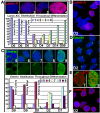
Figure 1. Differentiating hESC exhibit sweeping epigenetic and morphological changes
(A&B left) Small (5um) tightly packed round cells are characteristic of undifferentiated hESC. They stain brightly for pluripotency markers such as Alkaline Phosphatase (AP) (B, Left, red) (A+B Right) Differentiating cells peripheral to the colonies exhibit diminished expression of pluripotency markers (AP) and changes in nuclear size and shape. (C & Graph) SC35 (red) domain formation occurs as cells differentiate; either peripheral to the colonies in D0 cultures or with directed differentiation. (C1) Undifferentiated hESC show diffuse SC35. (C2-C3) SC35 domain formation starts as cells begin to differentiate. (C4) Mature SC35 domains, typical of somatic cells, are not abundant until D7 of differentiation. (D) Cajal bodies are absent in undifferentiated cells (left), are found in some differentiating peripheral cells in D0 cultures, and most differentiated cells (D14 Neuronal).
Figure 2
Figure 2. Maturation of the nuclear envelope (NE) occurs during hESC differentiation
(A) Lamin A (red) is not expressed in undifferentiated hESC (A1). It initiates expression in patches during early differentiation (A3), and is abundantly expressed throughout the cell by D4-7 (A4). (B) Initiation of lamin A often coincides with nuclear shape change. (C) Emerin (green) appears cytoplasmic in undifferentiated hESC (C5) but then declines (C6). Emerin localization to the NE also starts in patches concurrent with localization of Lamin A (C7) and is seen abundantly expressed by D4-7 (C8-9). (D) A field of “Patchy” localized Emerin in differentiating hESC. (E) Patchy emerin is usually seen in cells exhibiting undefined SC35 domains (cell from C7), while abundant emerin expression is usually seen in cells with clearly defined SC35 domains (cell from C9). (F) Lamin B is always present in undifferentiated 0 day hESC.
Figure 3
Figure 3. Distinct transformations in nuclear size and shape occur during a brief window of early differentiation
DNA staining reveals dramatic nuclear size and shape changes, in which occur often in synchronous waves of differentiating hES cells. (A)Undifferentiated cells in close proximity to each other within colonies (bottom) have smaller more uniform nuclei. Nuclear shape changes are prevalent in less densely packed areas of “differentiated” cells at the outer edge of the colony (top) (B) Moving away from the origin of the colony, nuclear size and shape change increases significantly in cells peripheral to the colony.
Figure 4
Figure 4. hES cells exhibit unusual PML structures
(A) Large unique linear (left) and rosette (middle) PML structures (red) are found in hESC cultures. Torus structures (right) are also seen, similar in shape to somatic PML bodies, but approximately 2-3 fold larger. (B) Unique PML structures are less abundant within undifferentiated colonies, and increase in abundance toward the colony’s edge and in differentiating cells outside the colony (see also E). The frequency decreases quickly upon differentiation, while the large torus structures peak later in differentiation. (C-D) Undifferentiated hESC (H9) also contain more somatic-like PML (C red, D green) structures, however they are much fewer in number per cell, and their numbers increase with differentiation (Tig-1 are somatic fibroblasts). (F) Unique PML structures are occasionally found in cells that still stain brightly for pluripotency markers (AP, green). (G) They are usually found in hES cells (top) lacking heterchromatin (HP1, green), which is found in cells containing more somatic-type PML bodies (bottom). Multiple examples of linear PML structures are shown (H), Linear Structures appear taut and can range in size from 2-10um. (I) PML Rosette structures can range in size from 2-8um in diameter and resemble “looping” threads or a honey-comb. (J) Both Linear and Rosette Structures appear to dissolve forming multiple small round PML structures.
Figure 5
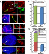
Figure 5. Unique PML structures exhibit a relationship to heterochromatin
(A) Linear PML Structures (green) often exhibit a relationship to centromeres (alpha satellite, red), at one or both ends of the structure, and appear “taut”. (B) Rosette PML structures also commonly encircle one or more centromeres. (C) The small round “somatic-like” PML structures in hESC also associate with centromeres, which is 2.5 fold higher than normal PML bodies in somatic cells. (D) Centromere interaction along the linear PML structure mapped predominantly to the ends suggesting a non random association. (E) Some linear PML structures maintained an association to chromatin (dapi, white) during mitosis. (F) PML structures associate with PCNA foci (red) in late S-phase cells, supporting an association to late replicating heterochromatin.
Figure 6
Figure 6. Interaction of Unique PML structures with the nuclear lamina
(A) Unique PML structures (green) frequently show a striking relationship with the nuclear lamina (red), either at the nuclear periphery (B 1st arrow) or within laminar folds as the cells undergo shape change (arrow A or 2nd arrow B). (B) PML structures often fill gaps in the lamina signal (see arrows in separated channels at right). (C) Approximately 52% of differentiated cells (D2) with Unique PML structures showed an interaction with Lamin A. (D) Lamin A expression begins focally at the NE of differentiatining cells. Unique PML structures are frequently found within these large foci. (E) Most hES cells containing a singular Lamin foci (75%) showed a close relationship with Unique PML structures.

References

    1. Ascoli CA, Maul GG. Identification of a novel nuclear domain. J.Cell Biol. 1991;112:785–795. - PMC - PubMed
    1. Bartova E, Galiova G, Krejci J, Harnicarova A, Strasak L, Kozubek S. Epigenome and chromatin structure in human embryonic stem cells undergoing differentiation. Dev Dyn. 2008a;237:3690–702. - PubMed
    1. Bartova E, Krejci J, Harnicarova A, Kozubek S. Differentiation of human embryonic stem cells induces condensation of chromosome territories and formation of heterochromatin protein 1 foci. Differentiation. 2008b;76:24–32. - PubMed
    1. Becker KA, Ghule PN, Therrien JA, Lian JB, Stein JL, van Wijnen AJ, Stein GS. Self-renewal of human embryonic stem cells is supported by a shortened G1 cell cycle phase. J Cell Physiol. 2006;209:883–93. - PubMed
    1. Bernardi R, Pandolfi PP. Structure, dynamics and functions of promyelocytic leukaemia nuclear bodies. Nat Rev Mol Cell Biol. 2007;8:1006–16. - PubMed

Publication types

MeSH terms

LinkOut - more resources