

Probing anomalous structural features in polypurine tract-containing RNA-DNA hybrids with neomycin B
- PMID: 19449839
- PMCID: PMC2802280
- DOI: 10.1021/bi900357j
Probing anomalous structural features in polypurine tract-containing RNA-DNA hybrids with neomycin B
Abstract
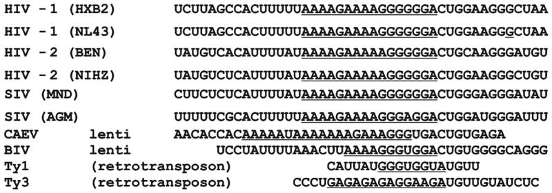
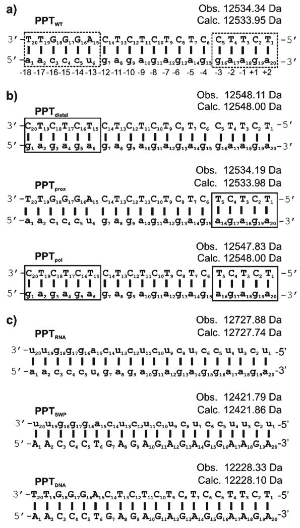
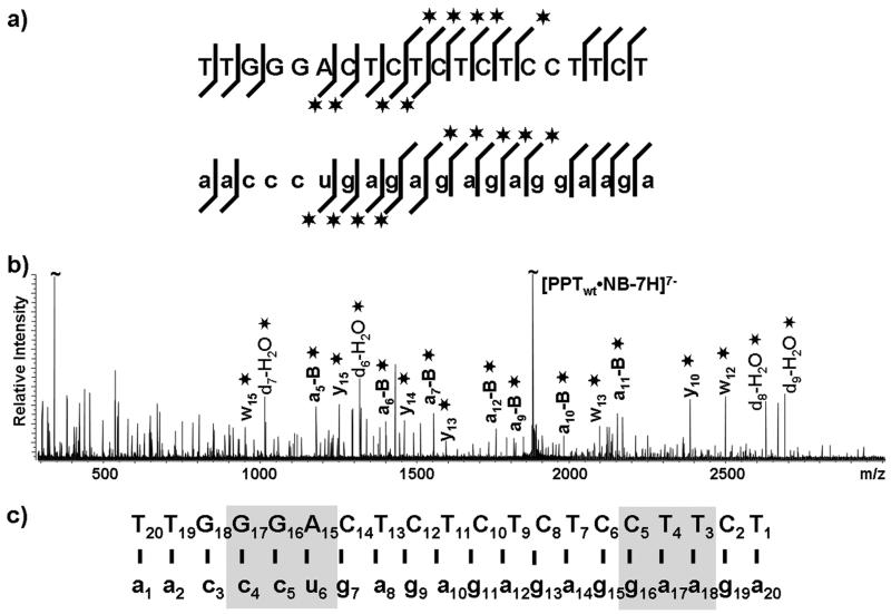
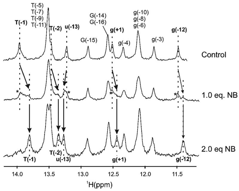
During (-)-strand DNA synthesis in retroviruses and Saccharomyces cerevisiae LTR retrotransposons, a purine rich region of the RNA template, known as the polypurine tract (PPT), is resistant to RNase H-mediated hydrolysis and subsequently serves as a primer for (+)-strand, DNA-dependent DNA synthesis. Although HIV-1 and Ty3 PPT sequences share no sequence similarity beyond the fact that both include runs of purine ribonucleotides, it has been suggested that these PPTs are processed by their cognate reverse transcriptases (RTs) through a common molecular mechanism. Here, we have used the aminoglycoside neomycin B (NB) to examine which structural features of the Ty3 PPT contribute to specific recognition and processing by its cognate RT. Using high-resolution NMR, direct infusion FTICR mass spectrometry, and isothermal titration calorimetry, we show that NB binds preferentially and selectively adjacent to the Ty3 3' PPT-U3 cleavage junction and in an upstream 5' region where the thumb subdomain of Ty3 RT putatively grips the substrate. Regions highlighted by NB on the Ty3 PPT are similar to those previously identified on the HIV-1 PPT sequence that are implicated as contact points for substrate binding by its RT. Our findings thus support the notion that common structural features of lentiviral and LTR-retrotransposon PPTs facilitate the interaction with their cognate RT.
Figures
Similar articles
-
Nonpolar thymine isosteres in the Ty3 polypurine tract DNA template modulate processing and provide a model for its recognition by Ty3 reverse transcriptase.J Biol Chem. 2003 Jul 18;278(29):26526-32. doi: 10.1074/jbc.M302374200. Epub 2003 Apr 30. J Biol Chem. 2003. PMID: 12730227
-
Structural probing of the HIV-1 polypurine tract RNA:DNA hybrid using classic nucleic acid ligands.Nucleic Acids Res. 2008 May;36(8):2799-810. doi: 10.1093/nar/gkn129. Epub 2008 Apr 9. Nucleic Acids Res. 2008. PMID: 18400780 Free PMC article.
-
Two modes of HIV-1 polypurine tract cleavage are affected by introducing locked nucleic acid analogs into the (-) DNA template.J Biol Chem. 2004 Aug 27;279(35):37095-102. doi: 10.1074/jbc.M403306200. Epub 2004 Jun 25. J Biol Chem. 2004. PMID: 15220330
-
Reverse Transcription in the Saccharomyces cerevisiae Long-Terminal Repeat Retrotransposon Ty3.Viruses. 2017 Mar 15;9(3):44. doi: 10.3390/v9030044. Viruses. 2017. PMID: 28294975 Free PMC article. Review.
-
'Binding, bending and bonding': polypurine tract-primed initiation of plus-strand DNA synthesis in human immunodeficiency virus.Int J Biochem Cell Biol. 2004 Sep;36(9):1752-66. doi: 10.1016/j.biocel.2004.02.016. Int J Biochem Cell Biol. 2004. PMID: 15183342 Review.
Cited by
-
Targeting the HIV RNA genome: high-hanging fruit only needs a longer ladder.Curr Top Microbiol Immunol. 2015;389:147-69. doi: 10.1007/82_2015_434. Curr Top Microbiol Immunol. 2015. PMID: 25735922 Free PMC article. Review.
-
Viral reverse transcriptases show selective high affinity binding to DNA-DNA primer-templates that resemble the polypurine tract.PLoS One. 2012;7(7):e41712. doi: 10.1371/journal.pone.0041712. Epub 2012 Jul 27. PLoS One. 2012. PMID: 22848574 Free PMC article.
-
Ty3 reverse transcriptase complexed with an RNA-DNA hybrid shows structural and functional asymmetry.Nat Struct Mol Biol. 2014 Apr;21(4):389-96. doi: 10.1038/nsmb.2785. Epub 2014 Mar 9. Nat Struct Mol Biol. 2014. PMID: 24608367 Free PMC article.
-
Synthesis and incorporation of 13C-labeled DNA building blocks to probe structural dynamics of DNA by NMR.Nucleic Acids Res. 2017 Sep 6;45(15):9178-9192. doi: 10.1093/nar/gkx592. Nucleic Acids Res. 2017. PMID: 28911104 Free PMC article.
-
New insights into transcription fidelity: thermal stability of non-canonical structures in template DNA regulates transcriptional arrest, pause, and slippage.PLoS One. 2014 Mar 3;9(3):e90580. doi: 10.1371/journal.pone.0090580. eCollection 2014. PLoS One. 2014. PMID: 24594642 Free PMC article.
References
-
- Champoux JJ. Roles of ribonuclease H in reverse transcription. In: Skalka AM, Goff SP, editors. Reverse Transcriptase. Cold Spring Harbor Laboratory Press; Plainview, NY: 1993. pp. 103–118.
-
- Telesnitsky A, Goff SP. Reverse transcriptase and the generation of retroviral DNA. Cold Spring Harbor Laboratory Press; Plainview, NY: 1997. - PubMed
-
- Rausch JW, Le Grice SF. ‘Binding, bending and bonding’: Polypurine tract-primed initiation of plus-strand DNA synthesis in human immunodeficiency virus. Int. J. Biochem. Cell Biol. 2004;36:1752–1766. - PubMed
-
- Bibillo A, Lener D, Tewari A, Le Grice SF. Interaction of the Ty3 reverse transcriptase thumb subdomain with template-primer. J. Biol. Chem. 2005;280:30282–30290. - PubMed
Publication types
MeSH terms
Substances
Grants and funding
LinkOut - more resources
Full Text Sources
Molecular Biology Databases
