

Mitochondrial death effectors: relevance to sarcopenia and disuse muscle atrophy
- PMID: 19450666
- PMCID: PMC2826514
- DOI: 10.1016/j.bbagen.2009.05.007
Mitochondrial death effectors: relevance to sarcopenia and disuse muscle atrophy
Abstract
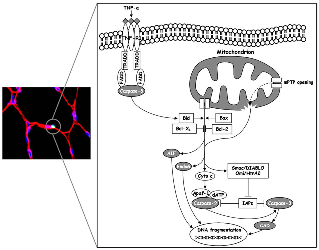
Accelerated apoptosis in skeletal muscle is increasingly recognized as a potential mechanism contributing to the development of sarcopenia of aging and disuse muscle atrophy. Given their central role in the regulation of apoptosis, mitochondria are regarded as key players in the pathogenesis of myocyte loss during aging and other atrophying conditions. Oxidative damage to mitochondrial constituents, impaired respiration and altered mitochondrial turnover have been proposed as potential triggering events for mitochondrial apoptotic signaling. In addition, iron accumulation within mitochondria may enhance the susceptibility to apoptosis during the development of sarcopenia and possibly acute muscle atrophy, likely through exacerbation of oxidative stress. Mitochondria can induce myocyte apoptosis via both caspase-dependent and independent pathways, although the apoptogenic mediators involved may be different depending on age, muscle type and specific atrophying conditions. Despite the considerable advances made, additional research is necessary to establish a definite causal link between apoptotic signaling and the development of sarcopenia and acute atrophy. Furthermore, a translational effort is required to determine the role played by apoptosis in the pathogenesis of sarcopenia and disuse-induced muscle loss in human subjects.
Published by Elsevier B.V.
Figures
References
-
- Roubenoff R. Sarcopenia: a major modifiable cause of frailty in the elderly. J. Nutr. Health Aging. 2000;4:140–142. - PubMed
-
- Rantanen T, Guralnik JM, Foley D, Masaki K, Leveille S, Curb JD, White L. Midlife hand grip strength as a predictor of old age disability. JAMA. 1999;281:558–560. - PubMed
-
- Metter EJ, Talbot LA, Schrager M, Conwit R. Skeletal muscle strength as a predictor of all-cause mortality in healthy men. J. Gerontol. A, Biol. Sci. Med. Sci. 2002;57:B359–B365. - PubMed
-
- Janssen I, Shepard DS, Katzmarzyk PT, Roubenoff R. The healthcare costs of sarcopenia in the United States. J. Am. Geriatr. Soc. 2004;52:80–85. - PubMed
Publication types
MeSH terms
Substances
Grants and funding
LinkOut - more resources
Full Text Sources
Other Literature Sources
Medical
