. 2009 May 15;23(10):1177-82.
doi: 10.1101/gad.511109.

Histone demethylase JMJD3 contributes to epigenetic control of INK4a/ARF by oncogenic RAS

Affiliations

Histone demethylase JMJD3 contributes to epigenetic control of INK4a/ARF by oncogenic RAS

Marta Barradas et al. Genes Dev. .

Abstract

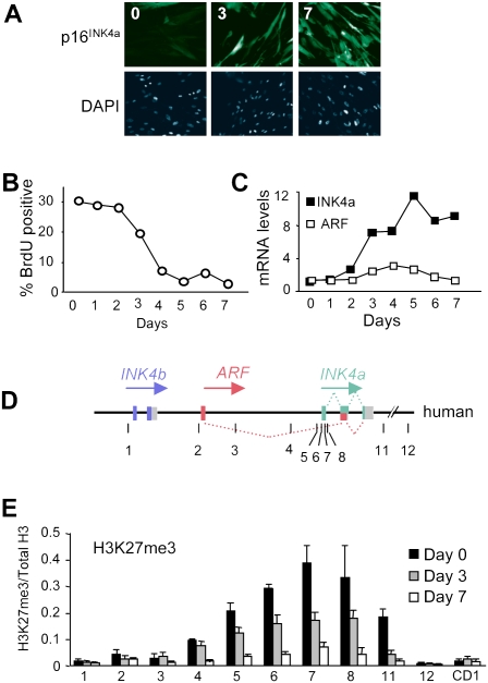
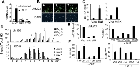
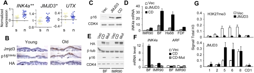
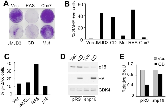
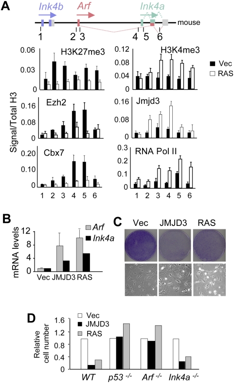
The INK4a/ARF tumor suppressor locus, a key executor of cellular senescence, is regulated by members of the Polycomb group (PcG) of transcriptional repressors. Here we show that signaling from oncogenic RAS overrides PcG-mediated repression of INK4a by activating the H3K27 demethylase JMJD3 and down-regulating the methyltransferase EZH2. In human fibroblasts, JMJD3 activates INK4a, but not ARF, and causes p16(INK4a)-dependent arrest. In mouse embryo fibroblasts, Jmjd3 activates both Ink4a and Arf and elicits a p53-dependent arrest, echoing the effects of RAS in this system. Our findings directly implicate JMJD3 in the regulation of INK4a/ARF during oncogene-induced senescence and suggest that JMJD3 has the capacity to act as a tumor suppressor.


Figures

Figure 1.
Figure 1.
Loss of H3K27me3 at the INK4a/ARF locus in cells undergoing OIS. (A) Induction of senescence and p16INK4a expression in IMR90-ER:RAS cells treated with OHT for 0, 3, and 7 d. (B,C) Percentage of BrdU-positive cells and relative levels of INK4a and ARF mRNAs as measured by qRT–PCR in IMR90-ER:RAS cells treated with OHT as indicated. The results are representative of three independent experiments. (D) Schematic representation of the human INK4b–ARF–INK4a locus (not to scale). Numbered lines show the approximate location of primer sets used for ChIP (Supplemental Table S1). (E) Degree of H3K27me3 modification at the INK4b–ARF–INK4a locus in IMR90-ER:RAS cells treated with OHT for 0, 3, and 7 d. A primer set amplifying the cyclin D1 gene (CD1) was used as a negative control. The results are presented as the ratio of H3K27me3 to total H3 as judged by ChIP analyses.
Figure 2.
Figure 2.
Up-regulation of JMJD3 by oncogenic RAS. (A) qRT–PCR assessment of the relative levels of JMJD3, UTX, and EZH2 mRNAs in IMR90-ER:RAS cells treated with OHT for 7 d normalized to untreated cells. (B) Increased levels of endogenous JMJD3 following induction of RAS. Nuclear staining using DAPI is shown in the bottom panel. (C) Increased expression of JMJD3 mRNA in IMR90 cells transduced with constitutively active forms of RAF or MEK1. (D) ChIP analyses showing enhanced binding of JMJD3 and reduced binding of EZH2 at the INK4a/ARF locus in IMR90-ER:RAS cells. (E, left) Knockdown of JMJD3 mRNA with two shRNAs. Controls and shRNAs were used to infect IMR90-ER:RAS cells. (Right) After selection, cells were treated with OHT for 48 h and pulsed with BrdU for 16 h. These results are representative of three independent experiments. (F) IMR90-ER:RAS cells were transfected with siRNAs or controls. Two days after, cells were treated with OHT for four more days, fixed, and subjected to immunofluorescence. Percentages of JMJD3-positive (left) or p16INK4a-positive (right) cells are shown.
Figure 3.
Figure 3.
Ectopic expression of JMJD3 activates p16INK4a in HDFs. (A) Expression of INK4a, JMJD3, and UTX in melanocytic nevi versus normal skin from Talantov et al. (2005). (**) P < 0.001; (*) P < 0.05. The P-values correspond to a nonparametric unpaired t-test. (B) Immunohistochemistry of Jmjd3 and p16Ink4a proteins in paraffin-embedded sections of mouse back skin. HA staining was used as a negative control. Similar results were obtained in mouse tail skin (not shown). (C) Immunoblot of p16INK4a in Hs68 fibroblasts transduced with JMJD3 or the CD construct. CDK4 was used as a loading control. (D) qRT–PCR showing that expression of full-length JMJD3 or the CD up-regulates INK4a in the indicated strains of the HDFs. (E) Immunoblot showing up-regulation of p16INK4a by the HA-tagged CD of JMJD3 (CD) but not by a catalytically inactive mutant (Mut). Equivalent results were obtained in BF and IMR90 cells. (F) Ectopic expression of JMJD3 CD in BF and IMR90 cells up-regulates INK4a but not ARF. (G) Expression of JMJD3 reduces the extent of H3K27me3 modification and increases the density of JMJD3 at the INK4a locus in IMR90 cells.
Figure 4.
Figure 4.
JMJD3 causes p16INK4a-dependent growth arrest in HDFs. (A) IMR90 cells were infected with the indicated retroviruses. Crystal violet staining at 10–15 d post-infection. (B) JMJD3, CD, and activated RAS cause the appearance of SAHFs, whereas Cbx7 delays senescence in HDFs. Quantification was performed on at least 200 nuclei for each. (C) Immunofluorescence for γH2AX indicates that RAS causes a DNA damage response in IMR90 cells, whereas JMJD3 or p16INK4a do not. The results presented in A–C are representative of three independent experiments. (D,E) shRNA knockdown of p16INK4a alleviates the arrest caused by ectopic JMJD3. Immunoblot showing p16INK4a and HA-CD. BrdU incorporation was quantified by immunofluorescence and relative numbers were plotted. Two independent experiments showed similar results.
Figure 5.
Figure 5.
In MEFs, JMJD3 activates both Arf and Ink4a, causing Arf-dependent growth arrest. (A) MEFs were infected with a control vector or with activated RAS. After selection, cells were subjected to ChIP using the indicated antibodies and primer sets (Supplemental Table S1). The approximate location of the primer sets used is shown. (B) In normal MEFs, ectopic expression of JMJD3 or RAS causes up-regulation of both Arf and Ink4a transcripts. (C) Ectopic expression of JMJD3 or RAS in MEFs causes a senescence-like arrest. The top panel shows crystal violet staining at 10–15 d post-infection. The bottom panel shows typical senescence-like morphology. (D) JMJD3 and RAS cause growth arrest in normal and Ink4a−/− MEFs but not in p53−/− or Arf−/− MEFs. Relative cell numbers were assessed by crystal violet staining. The data are representative of between two and six independent experiments for each genotype.

References

    1. Acosta JC, O'Loghlen A, Banito A, Guijarro MV, Augert A, Raguz S, Fumagalli M, Da Costa M, Brown C, Popov N, et al. Chemokine signaling via the CXCR2 receptor reinforces senescence. Cell. 2008;133:1006–1018. - PubMed
    1. Agger K, Cloos PA, Christensen J, Pasini D, Rose S, Rappsilber J, Issaeva I, Canaani E, Salcini AE, Helin K. UTX and JMJD3 are histone H3K27 demethylases involved in HOX gene regulation and development. Nature. 2007;449:731–734. - PubMed
    1. Agger K, Cloos PAC, Rudkjær L, Williams K, Andersen G, Christensen J, Helin K. The H3K27me3 demethylase JMJD3 contributes to the activation of the INK4A–ARF locus in response to oncogene- and stress-induced senescence. Genes & Dev. 2009 doi: 10.1101/gad.510809. (this issue) - DOI - PMC - PubMed
    1. Ananthanarayanan M, Li S, Balasubramaniyan N, Suchy FJ, Walsh MJ. Ligand-dependent activation of the farnesoid X-receptor directs arginine methylation of histone H3 by CARM1. J Biol Chem. 2004;279:54348–54357. - PubMed
    1. Bracken AP, Pasini D, Capra M, Prosperini E, Colli E, Helin K. EZH2 is downstream of the pRB–E2F pathway, essential for proliferation and amplified in cancer. EMBO J. 2003;22:5323–5335. - PMC - PubMed

Publication types

Substances