. 2009 May 15;23(10):1195-206.
doi: 10.1101/gad.512009.

The PRC1 Polycomb group complex interacts with PLZF/RARA to mediate leukemic transformation

Affiliations

The PRC1 Polycomb group complex interacts with PLZF/RARA to mediate leukemic transformation

Hanane Boukarabila et al. Genes Dev. .

Abstract

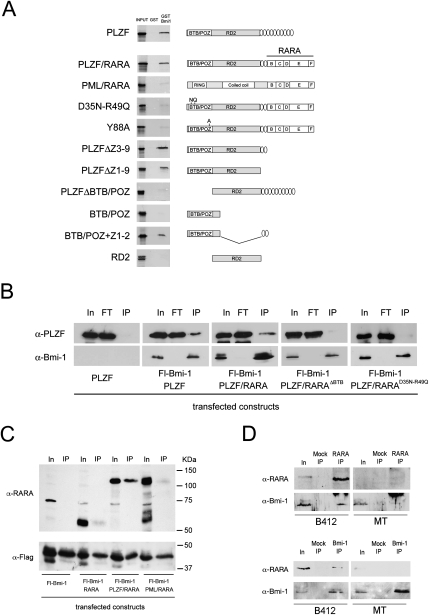
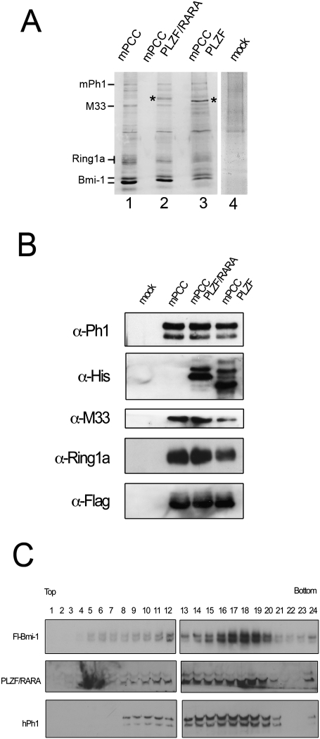
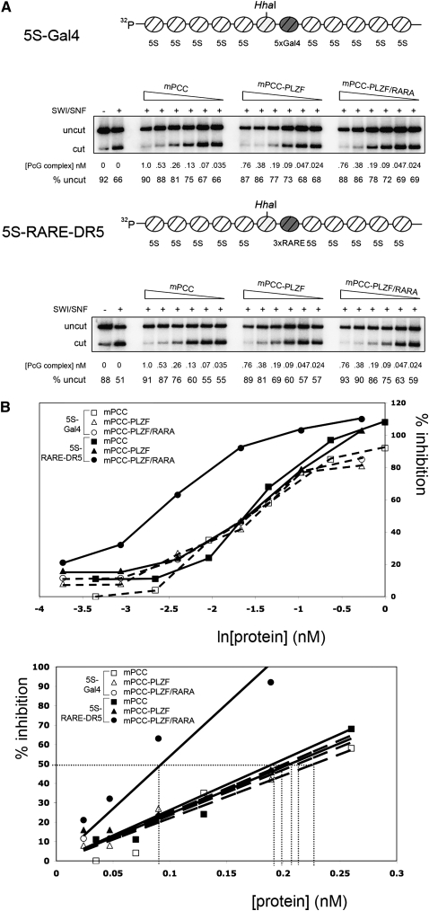
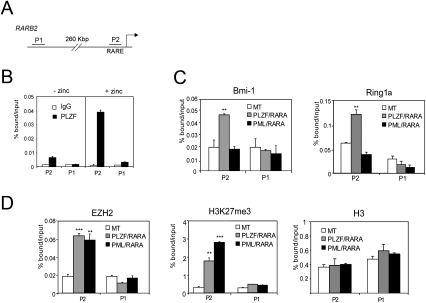
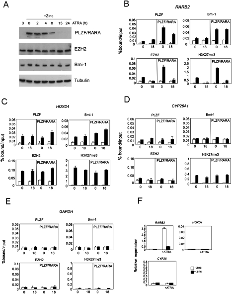
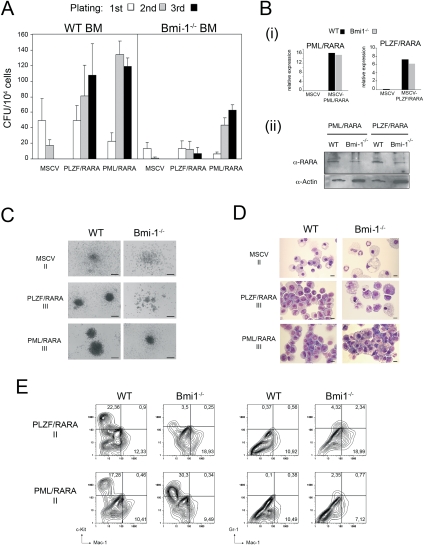
Ectopic repression of retinoic acid (RA) receptor target genes by PML/RARA and PLZF/RARA fusion proteins through aberrant recruitment of nuclear corepressor complexes drives cellular transformation and acute promyelocytic leukemia (APL) development. In the case of PML/RARA, this repression can be reversed through treatment with all-trans RA (ATRA), leading to leukemic remission. However, PLZF/RARA ectopic repression is insensitive to ATRA, resulting in persistence of the leukemic diseased state after treatment, a phenomenon that is still poorly understood. Here we show that, like PML/RARA, PLZF/RARA expression leads to recruitment of the Polycomb-repressive complex 2 (PRC2) Polycomb group (PcG) complex to RA response elements. However, unlike PML/RARA, PLZF/RARA directly interacts with the PcG protein Bmi-1 and forms a stable component of the PRC1 PcG complex, resulting in PLZF/RARA-dependent ectopic recruitment of PRC1 to RA response elements. Upon treatment with ATRA, ectopic recruitment of PRC2 by either PML/RARA or PLZF/RARA is lost, whereas PRC1 recruited by PLZF/RARA remains, resulting in persistent RA-insensitive gene repression. We further show that Bmi-1 is essential for the PLZF/RARA cellular transformation property and implicates a central role for PRC1 in PLZF/RARA-mediated myeloid leukemic development.


Figures

Figure 1.
Figure 1.
PLZF/RARA interacts with Bmi-1 through its BTB-POZ domain. (A) GST and GST-Bmi-1 pull-down of 35S-labeled wild-type and mutant isoforms of PLZF, PLZF/RARA, and PML/RARA. Structures of PLZF, PLZF/RARA, and PML/RARA isoforms used in GST pull-down and co-IP experiments are shown. (B) Nuclei of 293T cells cotransfected with Flag-Bmi-1 and the PLZF or PLZF/RARA constructs were immunoprecipitated with anti-Flag beads and were immunoblotted with either an anti-PLZF or anti-Bmi-1 antibody. (IP) 5% of the input; (FT) the flow-through. (C) Nuclei of 293T cells cotransfected with Fl-Bmi-1 and RARA, PLZF/RARA, or PML/RARA were immunoprecipitated with anti-Flag beads and immunoblotted with either an anti-RARA or anti-Flag antibody. (IP) 2.5% of the input. (D) Nuclei of U937-MT (control) of U937-B412 (PLZF/RARA) cells pretreated with zinc were immunoprecipitated with anti-RARA (top panels) or anti-Bmi-1 antibodies (bottom panels) and immunoblotted for PLZF/RARA (α-RARA) or Bmi-1 (α-Bmi-1). Mock IP represents immunoprecipitation without antibody; RARA IP and Bmi-1 IP represent 2.5% of input.
Figure 2.
Figure 2.
PLZF and PLZF/RARA associate with the reconstituted PRC1 complex. (A) Colloidal Coomassie blue-stained gel of purified mPCC (lane 1), mPCC–PLZF/RARA (lane 2), mPCC–PLZF (lane 3), and mock (lane 4) complexes. Asterisks highlight the bands corresponding to PLZF/RARA (lane 2) and PLZF (lane 3) verified by Western analyses. The mock complex corresponds to anti-Flag purification of extracts from Sf9 cells infected with mPh1, PLZF/RARA, M33, and Ring1a viruses. Densitometry analyses of the Coomassie-stained complexes show that the various mPCC complexes are ∼55%–65% pure, when compared with the mock purified complex. (B) Western blots of the complexes shown in A. PLZF/RARA and PLZF were detected by an anti-His tag antibody, and Bmi-1 was revealed by an anti-Flag tag antibody. (C) Western analyses of the sedimentation profiles of Bmi-1, PLZF/RARA, and mPh1 following fraction of the mPCC–PLZF/RARA complex by glycerol gradient sedimentation, showing that PLZF/RARA remains associated with the mPCC complex.
Figure 3.
Figure 3.
PLZF/RARA recruits PRC1 to chromatin containing RARE elements. (A) REA assays on in vitro reconstituted 5S chromatin arrays. PhosphorImager scans of HhaI digestion products following chromatin incubation in the presence or absence of SWI/SNF and PCC complexes with the 5S-Gal4 template (top panel) or the 5S-RARE-DR5 template (bottom panel). Percentages of template undigested by HhaI are given. The concentrations of PCC complexes (in nanomolar) used in the reactions are the measured concentration of active DNA-binding molecules. Schematic representation of 5S array templates used in the REA assays are shown above the data. “5S-Gal4” contains five Gal4-binding sites downstream from the unique HhaI restriction site, whereas “5S-RARE-DR5” contains three RARE DR5 elements. (B) Data from REA assays shown in A are graphically represented as percentage of inhibition (Francis et al. 2001) as a function of ln (active protein concentration) (top graph) and active protein concentration (bottom graph), showing increased efficiency of the PCC–PLZF/RARA complex to inhibit chromatin remodeling when RARE DR5 elements are present in the chromatin. (Bottom graph) The linear section of the data is shown, where the quantities of active complex required to achieve 50% inhibition of chromatin remodeling are highlighted (dotted line), demonstrating that PCC–PLZF/RARA is approximately twofold more efficient at inhibition of chromatin remodeling than PCC and PCC–PLZF when RARE DR5 elements are present.
Figure 4.
Figure 4.
PLZF/RARA targets PRC1 and PRC2 to RARβ2 promoter. (A) Representation of the RARβ2 promoter. P1 and P2 indicate the regions amplified by PCR. (B) PLZF/RARA on RARβ2 promoter was analyzed by quantitative ChIP (qChIP) with an anti-PLZF antibody on chromatin fragments prepared from U937-B412 cells pretreated with zinc or untreated. (**) P < 0.01 (C,D) qChIP analyses of the PRC1 subunits Bmi-1 and Ring1a (C), or EZH2, H3K27me3, and histone H3 (D) at RARβ2 promoter in the absence (MT) or in the presence of either PLZF/RARA or PML/RARA. Error bars represent standard deviations obtained from at least two independent experiments. (**) P < 0.01; (***) P < 0.001.
Figure 5.
Figure 5.
Effect of ATRA treatment on PLZF/RARA-mediated PRC1 and PRC2 recruitment. (A) Western analyses of nuclear extracts prepared from PLZF/RARA-expressing cells (+Zinc) at various times of ATRA treatment. Proteins were blotted and probed with antibodies to PLZF, EZH2, Bmi-1, and β-tubulin. (B–E) Analyses of the effects no stimulation (0) or 18 h of ATRA stimulation (18) on the enrichment of PLZF/RARA, Bmi-1, EZH2, and H3K27me3 in untreated B412 cells or B412 cells expressing PLZF/RARA (PLZF/RARA panels) at the RARβ2 promoter (B), the HOXD4 promoter (C), the CYP26A1 promoter (D), or the GAPDH promoter (E). Data are presented as percentage of bound/input and error bars indicate the standard deviation obtained from two or three independent experiments. White bars represent mock immunoprecipitation; black bars represent specific immunoprecipitation with the indicated antibodies. (F) The expression levels of RARβ2, HOXD4, and CYP26A1 were analyzed by quantitative real time PCR (qRT–PCR). RNA from U937-B412 cells (white bars) or zinc-pretreated U937-B412 cells (black bars; +zinc), either unstimulated or stimulated with 1 μM ATRA for 18 h (+ATRA) was extracted and 1–2 μg of RNA was reverse-transcribed. Gene expression of each gene is shown relative to HPRT expression.
Figure 6.
Figure 6.
Bmi-1 is required for PLZF/RARA transformation. (A) Bar charts indicate the number of colonies formed in methylcellulose cultures of lin BM transduced with the empty vector (MSCV), MSCV-PLZF/RARA (PLZF/RARA), or MSCV-PML/RARA (PML/RARA). Data represent the number of colonies from wild-type and Bmi-1−/− BM during three successive replatings. Error bars are standard deviation of the mean from five (PLZF/RARA) or two (PML/RARA) independent replating experiments. (B) Transgene expression was assessed by qRT-PCR (panel i) and immunoblotting (panel ii) from wild-type (WT) or Bmi-1−/− BM cells infected with either PLZF/RARA or PML/RARA viruses harvested after the third passage. Fusion proteins were detected in panel ii using antibodies against PLZF (PLZF/RARA) or RARA (PML/RARA). (C,D) Typical morphology (C) and MGG staining (D) of the second-round (II) or third-round (III) colonies generated from wild-type or Bmi-1−/− cells transduced with empty vector, PLZF/RARA, or PML/RARA. Bars: C, 200 μm; D, 10 μm. (E) Surface marker expression analyzed by FACS of the second-round colonies generated from wild-type or Bmi-1−/− cells transduced with PLZF/RARA or PML/RARA.

References

    1. Barna M, Merghoub T, Costoya JA, Ruggero D, Branford M, Bergia A, Samori B, Pandolfi PP. Plzf mediates transcriptional repression of HoxD gene expression through chromatin remodeling. Dev Cell. 2002;3:499–510. - PubMed
    1. Batsche E, Yaniv M, Muchardt C. The human SWI/SNF subunit Brm is a regulator of alternative splicing. Nat Struct Mol Biol. 2006;13:22–29. - PubMed
    1. Beisel C, Buness A, Roustan-Espinosa IM, Koch B, Schmitt S, Haas SA, Hild M, Katsuyama T, Paro R. Comparing active and repressed expression states of genes controlled by the Polycomb/Trithorax group proteins. Proc Natl Acad Sci. 2007;104:16615–16620. - PMC - PubMed
    1. Bel S, Coré N, Djabali M, Kieboom K, Van der Lugt N, Alkema MJ, Van Lohuizen M. Genetic interactions and dosage effects of Polycomb group genes in mice. Development. 1998;125:3543–3551. - PubMed
    1. Di Croce L, Raker VA, Corsaro M, Fazi F, Fanelli M, Faretta M, Fuks F, Lo Coco F, Kouzarides T, Nervi C, et al. Methyltransferase recruitment and DNA hypermethylation of target promoters by an oncogenic transcription factor. Science. 2002;295:1079–1082. - PubMed

Publication types

MeSH terms