. 2009 Jun 8;206(6):1365-78.
doi: 10.1084/jem.20090127. Epub 2009 May 18.

iNKT cell development is orchestrated by different branches of TGF-beta signaling

Affiliations

iNKT cell development is orchestrated by different branches of TGF-beta signaling

Jean-Marc Doisne et al. J Exp Med. .

Abstract

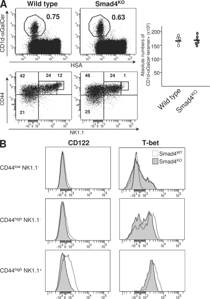
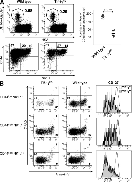
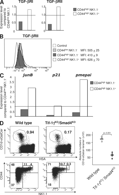
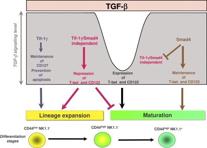
Invariant natural killer T (iNKT) cells constitute a distinct subset of T lymphocytes exhibiting important immune-regulatory functions. Although various steps of their differentiation have been well characterized, the factors controlling their development remain poorly documented. Here, we show that TGF-beta controls the differentiation program of iNKT cells. We demonstrate that TGF-beta signaling carefully and specifically orchestrates several steps of iNKT cell development. In vivo, this multifaceted role of TGF-beta involves the concerted action of different pathways of TGF-beta signaling. Whereas the Tif-1gamma branch controls lineage expansion, the Smad4 branch maintains the maturation stage that is initially repressed by a Tif-1gamma/Smad4-independent branch. Thus, these three different branches of TGF-beta signaling function in concert as complementary effectors, allowing TGF-beta to fine tune the iNKT cell differentiation program.


Figures

Figure 1.
Figure 1.
Functional thymic environment to select iNKT cells in the absence of TGF-β signaling. (A) Flow cytometric analysis of splenocytes in mixed BM chimeras. T cell–depleted BM from 2-wk-old CD4-Cre x TGF-βRIIfl/fl mice or wild-type littermate mice were mixed with BM from CD1dKO mice (ratio 1:1) and transferred into rag2KO animals. 6–7 wk later, the presence in the spleen of iNKT cells was investigated using CD1d-αGalCer tetramers and TCR-β staining. The results are representative of three different experiments with three animals per group. (B) Proliferation analysis of CD1d-restricted Vα14-expressing DN32D3 hybridoma in the presence of thymocytes from either wild-type animals or CD4-Cre x TGF-βRIIfl/fl mice and different concentration of αGalCer. Mean ± SD. n = 2, from two independent experiments.
Figure 2.
Figure 2.
Control of iNKT cell precursor development by TGF-β signaling. Flow cytometric analysis of the presence of iNKT cells in the thymus (A), liver (B), or spleen (C) of 14-d-old CD4-Cre x TGF-βRIIfl/fl mice (TGF-βRIIKO), CD4-Cre x Stopfl/fl TGF-βRICA mice (TGF-βRICA), or wild-type mice (TGF-βRIIwt). Absolute numbers of CD1d-αGalCer tetramer+ cells among either HSAlow cells or B220+ cells are illustrated by graphs and absolute numbers for each development stage are illustrated in supplemental table 1. CD44, NK1.1 staining on purified CD1d-αGalCer tetramers2 thymocytes (A) and on CD1d-αGalCer tetramers+ B220 cells from either the liver (B) or spleen (C). IL-4, IFN-γ, staining on either NK1.1 or NK1.1+ CD1d-αGalCer tetramer+ B220 cells from spleen (C). The results are representative of three or four different experiments with three animals per group.
Figure 3.
Figure 3.
Control iNKT cell precursor apoptosis by TGF-β signaling. Flow cytometric analysis of CD1d-αGalCer tetramer+ enriched from thymus from 14-d-old CD4-Cre x TGF-βRIIfl/fl mice (TGF-βRIIKO), CD4-Cre x Stopfl/fl TGF-βRICA mice (TGF-βRICA), or wild-type mice (TGF-βRIIwt) and stained for either CD44, NK1.1, or annexin V and incubated with 7-AAD (A) or CD44, NK1.1, and CD127 (B). These results are representative of three different experiments with two or three mice per group.
Figure 4.
Figure 4.
Control of T-bet and CD122 expression by TGF-β signaling. (A and B) Flow cytometric analysis of CD1d-αGalCer tetramer+ cells enriched from thymocytes from either CD4-Cre x TGF-βRIIfl/fl mice (TGF-βRIIKO; A) or CD4-Cre x Stopfl/fl TGF-βRICA mice (TGF-βRICA; B) stained for CD44, NK1.1, T-bet, and CD122. These results are representative of four different experiments with two mice per group.
Figure 5.
Figure 5.
Role of Smad4 signaling in iNKT cell differentiation. (A) Flow cytometric analysis of the presence of iNKT cells in the thymuses from either 16-d-old CD4-Cre x Smad4 mice (Smad4KO) or wild-type mice and staining for CD44 and NK1.1 on CD1d-αGalCer tetramer+ cells from the same thymuses. Absolute numbers of CD1d-αGalCer tetramer+ cells among either HSAlow cells are illustrated by graphs and absolute numbers for each development stage are illustrated in Table S1. (B) Flow cytometric analysis on CD1d-αGalCer tetramer+ cells enriched from thymuses from either CD4-Cre x Smad4fl/fl mice (Smad4KO) or wild-type mice littermate stained for CD44, NK1.1, T-bet, and CD122. These results are representative of three different experiments with two mice per group.
Figure 6.
Figure 6.
Role of Tif-1γ signaling in iNKT cell differentiation. (A) Flow cytometric analysis of the presence of iNKT cells in thymuses from either 16 d-old CD4-Cre x Tif-1γfl/fl mice (Tif-1γKO) or wild-type mice and staining for CD44 and NK1.1 on CD1d-αGalCer tetramer+ cells from the same thymuses. Absolute numbers of CD1d-αGalCer tetramer+ cells among either HSAlow cells are illustrated by graphs and absolute numbers for each development stage are illustrated in supplemental table 1. B) Flow cytometric analysis on CD1d-αGalCer tetramer+ cells enriched from thymuses from either CD4-Cre x Tif-1γfl/fl mice (Tif-1γKO) or wild-type littermate mice and stained for CD44, NK1.1, Annexin V, 7-AAD, and CD127. Mean fluorescence intensity is reported on the CD127 histograms. These results are representative of three different experiments with two mice per group.
Figure 7.
Figure 7.
Role of Tif-1γ/Smad4-independent signaling in the control of iNKT maturation. (A) tgf-b receptor expression analysis by RT-PCR performed on purified CD1d-αGalCer thymocytes from 16-d-old C57BL/6 mice. Relative gene expression are normalized on both hprt and g3pdh expression. (B) Flow cytometric analysis of CD1d-αGalCer tetramer+ cells from iNKT cell-enriched thymocytes of C57BL/6 mice, stained for TGF-βRII. Means of fluorescence intensity (MFI) are reported with their SD. (C) jun-b, p21, pme-pai expression analysis by RT-PCR performed on purified CD1d-αGalCer thymocytes from C57BL/6 mice. Relative gene expression was normalized on βactin expression. The results are representative of three different experiments from five to eight mice. (D) Flow cytometric analysis of the presence of iNKT cells in thymuses from either 13–14-d-old CD4-Cre x Tif-1γfl/fl x Smad4fl/fl mice or wild-type littermate mice and staining for CD44 and NK1.1 on CD1d-αGalCer tetramer+ cells from the same thymuses. Absolute numbers of CD1d-αGalCer tetramer+ cells among HSAlow cells are illustrated by graphs, and absolute numbers for each development stage are illustrated in Table S1. The results are representative of three different experiments with at least three mice per group.
Figure 8.
Figure 8.
Schematic model summarizing the effects of the three branches of TGF-β signaling on iNKT cell differentiation. The Tif-1γ–dependent branch prevents the apoptosis and allows lineage expansion. The Tif-1γ/Smad4-independent branch prevents maturation, and thus allows lineage expansion. The Smad4-dependent branch allows maturation.

References

    1. Kronenberg M., Gapin L. 2002. The unconventional lifestyle of NKT cells.Nat. Rev. Immunol. 2:557–568 - PubMed
    1. Kronenberg M., Rudensky A. 2005. Regulation of immunity by self-reactive T cells.Nature. 435:598–604 - PubMed
    1. Godfrey D.I., MacDonald H.R., Kronenberg M., Smyth M.J., Van Kaer L. 2004. NKT cells: what's in a name? Nat. Rev. Immunol. 4:231–237 - PubMed
    1. Kronenberg M. 2005. Toward an understanding of NKT cell biology: progress and paradoxes.Annu. Rev. Immunol. 23:877–900 - PubMed
    1. Bendelac A., Savage P.B., Teyton L. 2007. The biology of NKT cells.Annu. Rev. Immunol. 25:297–336 - PubMed

Publication types

MeSH terms

Substances