. 2009 May 18;185(4):577-86.
doi: 10.1083/jcb.200810035.

Heterochromatin protein 1 is recruited to various types of DNA damage

Affiliations

Heterochromatin protein 1 is recruited to various types of DNA damage

Martijn S Luijsterburg et al. J Cell Biol. .

Abstract

Heterochromatin protein 1 (HP1) family members are chromatin-associated proteins involved in transcription, replication, and chromatin organization. We show that HP1 isoforms HP1-alpha, HP1-beta, and HP1-gamma are recruited to ultraviolet (UV)-induced DNA damage and double-strand breaks (DSBs) in human cells. This response to DNA damage requires the chromo shadow domain of HP1 and is independent of H3K9 trimethylation and proteins that detect UV damage and DSBs. Loss of HP1 results in high sensitivity to UV light and ionizing radiation in the nematode Caenorhabditis elegans, indicating that HP1 proteins are essential components of DNA damage response (DDR) systems. Analysis of single and double HP1 mutants in nematodes suggests that HP1 homologues have both unique and overlapping functions in the DDR. Our results show that HP1 proteins are important for DNA repair and may function to reorganize chromatin in response to damage.


Figures
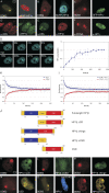
Figure 1.
Figure 1.
Recruitment of HP1s to UV damage depends on the CSD. (A–C, right) Damage-induced accumulation of mRFP–HP1-α (A), SCFP–HP1-β (B), and EGFP–HP1-γ (C) in living HeLa or MRC5 cells 30 min after local irradiation at 100 J/m2 though 5-µm pores. (A–C, left) The site of local DNA damage is indicated by accumulation of FP-tagged DDB2. (D–F, right) Immunolabeling of endogenous HP1-α (D), HP1-β (E), and HP1-γ (F) in locally UV-irradiated confluent human fibroblasts irradiated at 100 J/m2 through 3- (D and E) or 8-µm pores (F; cells are shown 30 min after irradiation). UV-damaged sites are visualized by local accumulation of XPA (D and E) or PCNA (F). (G) Accumulation of SCFP–HP1-β during the first 15 min after localized UV-C laser damage. Fig. S1 (B and C) shows the predamage distribution of HP1 in the same cell as shown in G and accumulation of DDB2-mCherry to indicate the site of damage. (H and I) Combined FLIP/FRAP analysis on NIH/3T3 cells expressing EGFP–HP1-β. Cells were either mock treated (H) or globally irradiated at 25 J/m2 (I). Half of a cell nucleus was bleached, and FLIP was measured in the nonbleached half 30 min after UV irradiation (blue line), whereas FRAP was measured in the bleached half (red line). (J) HP1-β deletion mutants. The CSD is indicated in red, the CD in blue, and the hinge in yellow. Numbers represent amino acid positions. (K–N) Nuclear localization of EGFP–HP1-β (ΔCD) (K), EGFP–HP1-β (ΔCSD) (L), EGFP–HP1-β (Δhinge) (M), and EYFP-CSD (N) in living HeLa or MRC5 cells locally irradiated at 100 J/m2 through 5-µm pores. (O and P) Recruitment of SCFP–HP1-β in Suv3-9–deficient mouse cells (O) and recruitment of EYFP–HP1-βT51A in MRC5 cells (P). The site of local DNA damage is indicated by accumulation of DDB2-mVenus or DDB2-mCherry, and images were taken 30 min after UV irradiation. Error bars indicate SD.
Figure 2.
Figure 2.
Recruitment of HP1 in NER-deficient cells. (A–F) Damage-induced accumulation of SCFP–HP1-β, EGFP–HP1-β, and EGFP–HP1-g in human fibroblasts deficient for XPA (A), XPC (B), DDB2 (C), MEFs deficient for XPC (D), human fibroblasts deficient for CSB (E), and human Seckel cells (F), which have severely reduced expression of ATR kinase. Cells were locally UV lamp irradiated at 100 J/m2 through 5-µm pores (A and C) or irradiated using a UV-C laser (B and D–F). The site of local DNA damage is indicated by accumulation of DDB2-mVenus, DDB2-mCherry, XPC-mVenus (A–C), or by a square (D and F). Images were taken 30 min after UV irradiation.
Figure 3.
Figure 3.
Long-term accumulation of HP1-β in repair-proficient and repair-deficient cells. (A) Repair-deficient XP-A cells were transfected with DDB2-mVenus and SCFP–HP1-β. Cells were irradiated at 100 J/m2, and accumulation of HP1-β was monitored for 4 h after UV irradiation. (B) Repair-deficient XP-A cells were transfected with mVenus-XPA (to complement the repair-deficient phenotype), DDB2-mCherry, and SCFP–HP1-β. Cells were irradiated at 100 J/m2, and accumulation of HP1-β was monitored for 4 h after UV irradiation. (C) Wild-type (MRC5) cells were transfected with DDB2-mCherry and YFP-CSD, locally irradiated (100 J/m2), and accumulation of the CSD was monitored for 4 h after UV irradiation. The accumulation of DDB2-mVenus or DDB2-mCherry indicates the site of local damage.
Figure 4.
Figure 4.
Recruitment of HP1-β to DSBs. (A) Mouse cells expressing EGFP–HP1-β (green) were locally irradiated with soft x rays through a nickel mask with pores of 5 µm and subsequently labeled for γH2AX (red). (B) Mouse cells expressing EGFP–HP1-β (green) were irradiated with α-particles and subsequently labeled for γH2AX (red). (C) Hamster cells deficient in Ku80 were irradiated with α-particles and subsequently labeled for endogenous HP1-β (green) and γH2AX (red). Cells are shown 30 min after irradiation. (D–I) Wild-type U2OS cells expressing various HP1-β fusion proteins were sensitized with 10 µg/ml Hoechst 33342 for 5 min and locally irradiated (five iterations) in a strip spanning the nucleus using a 405-nm laser at 70% output. GFP–HP1-β before and after laser-assisted damage (the damaged area is indicated by arrows; 5 min; D and E), GFP–HP1-βΔCD (F), YFP–HP1-βT51A (G), GFP–HP1-βΔCSD (H), and YFP-CSD (I). Accumulation of NBS1-mCherry indicates the site of laser-induced DNA damage. (J) GFP–HP1-β accumulation in BrdU-sensitized U2OS cells during the first 800 s after irradiation using a 337-nm laser. (K) Quantification of GFP–HP1-β accumulation as described in J. Error bars indicate SD.
Figure 5.
Figure 5.
Survival of C. elegans HP1 knockout worms upon UV irradiation and IR. (A) Hatching of wild-type, hpl-1Δ, hpl-2ts, and hpl-2ts/hpl-1Δ mutant eggs 8 h after collection. (B) Hatching of wild-type, hpl-1Δ, hpl-2ts, and hpl-2ts/hpl-1Δ mutant eggs 8 h after collection and subsequently irradiated with UV-B at 80 J/m2. (C) Quantification of hatching and nonhatching eggs after UV irradiation relative to nonirradiated eggs. In addition to wild-type, hpl-1Δ, hpl-2ts, and hpl-2ts/hpl-1Δ mutant eggs, the survival of xpa mutant eggs was also quantified. (D) Quantification of hatching and nonhatching eggs after IR (40–120 Gy) relative to nonirradiated eggs. Bars show the survival of wild-type eggs and of hpl-1Δ, hpl-2ts, and hpl-2ts/hpl-1Δ mutant eggs. Representative assays performed in duplicate or in quintuplicate are shown. For each assay, at least 120 animals were scored. Error bars indicate SD.

Comment in

Similar articles

Cited by

References

    1. Agarwal B.K., Sparrow J.H. 1981. Line intensities in the soft x-ray region.J. Phys. F Met. Phys. 11:1303–1309
    1. Aten J.A., Stap J., Krawczyk P.M., van Oven C.H., Hoebe R.A., Essers J., Kanaar R. 2004. Dynamics of DNA double-strand breaks revealed by clustering of damaged chromosome domains.Science. 303:92–95 - PubMed
    1. Aucott R., Bullwinkel J., Yu Y., Shi W., Billur M., Brown J.P., Menzel U., Kioussis D., Wang G., Reisert I., et al. 2008. HP1-β is required for development of the cerebral neocortex and neuromuscular junctions.J. Cell Biol. 183:597–606 - PMC - PubMed
    1. Ayoub N., Jeyasekharan A.D., Bernal J.A., Venkitaraman A.R. 2008. HP1-beta mobilization promotes chromatin changes that initiate the DNA damage response.Nature. 453:682–686 - PubMed
    1. Bartek J., Lukas J. 2007. DNA damage checkpoints: from initiation to recovery or adaptation.Curr. Opin. Cell Biol. 19:238–245 - PubMed

Publication types