. 2009 May 18;185(4):641-55.
doi: 10.1083/jcb.200812042.

TIP47 functions in the biogenesis of lipid droplets

Affiliations

TIP47 functions in the biogenesis of lipid droplets

Anna V Bulankina et al. J Cell Biol. .

Abstract

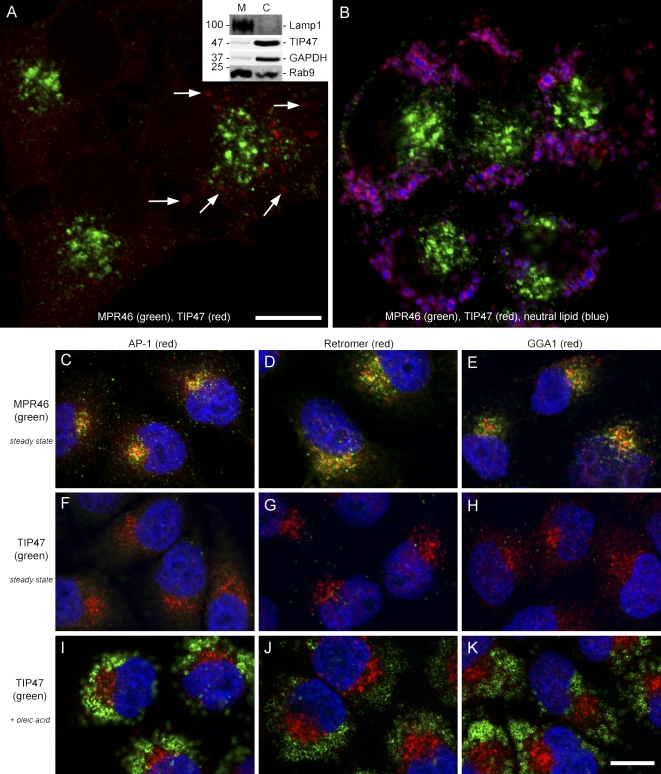
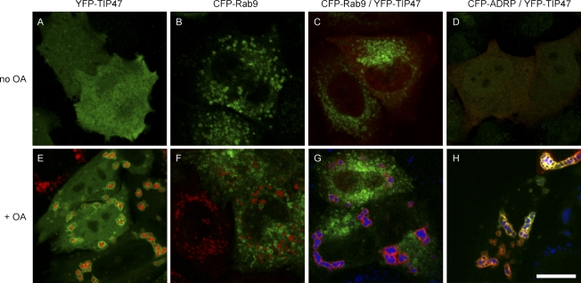
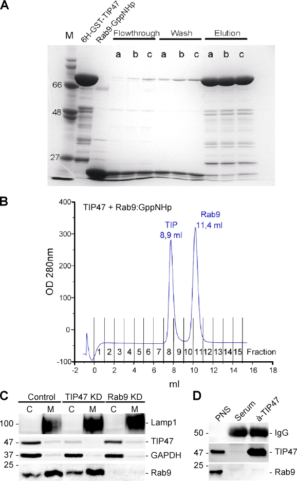
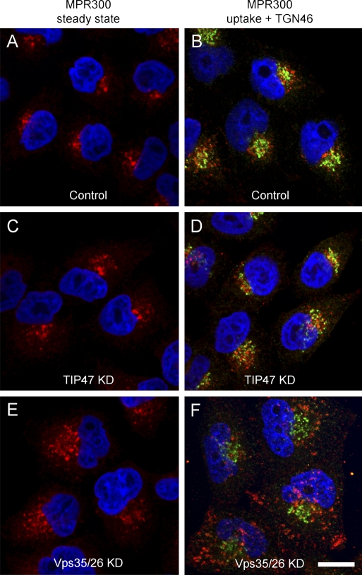
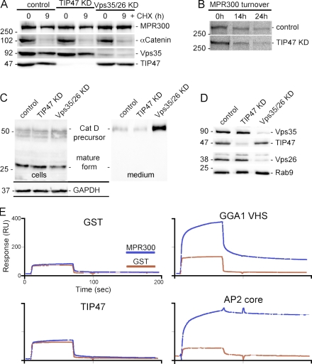
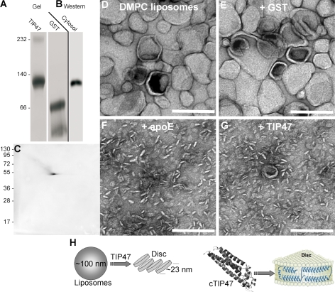
TIP47 (tail-interacting protein of 47 kD) was characterized as a cargo selection device for mannose 6-phosphate receptors (MPRs), directing their transport from endosomes to the trans-Golgi network. In contrast, our current analysis shows that cytosolic TIP47 is not recruited to organelles of the biosynthetic and endocytic pathways. Knockdown of TIP47 expression had no effect on MPR distribution or trafficking and did not affect lysosomal enzyme sorting. Therefore, our data argue against a function of TIP47 as a sorting device. Instead, TIP47 is recruited to lipid droplets (LDs) by an amino-terminal sequence comprising 11-mer repeats. We show that TIP47 has apolipoprotein-like properties and reorganizes liposomes into small lipid discs. Suppression of TIP47 blocked LD maturation and decreased the incorporation of triacylglycerol into LDs. We conclude that TIP47 functions in the biogenesis of LDs.


Figures

Figure 1.
Figure 1.
TIP47 and MPR46 localize to different compartments. (A and B) Staining of HeLa cells for MPR46 (green), TIP47 (red), and neutral lipid (blue) at steady state (A) or after oleic acid feeding (B). Arrows point to small TIP47-positive structures. (inset) Western blot for the indicated proteins after fractionation of cells into membranes (M) and cytosol (C). Values are shown in kilodaltons. (C–K) Staining of MPR46, TIP47, and the indicated sorting proteins in HeLa cells at steady state (C–H) or after oleic acid feeding (I–K). Colocalization is indicated in yellow. Bars, 15 µm.
Figure 2.
Figure 2.
Endogenous TIP47 and Rab9 do not colocalize. HeLa cells were fixed and stained for the indicated proteins. GFP-Rab7 and GFP-Lamp1 were transiently transfected 48 h prior to fixation. Colocalization is indicated in yellow. The inset displays a cell with small LDs at steady state. Bar, 15 µm.
Figure 3.
Figure 3.
CFP-Rab9 does not colocalize with YFP-TIP47. (A–H) HeLa cells were transiently transfected with YFP-TIP47 (A and E, green), CFP-Rab9 (B and F, green), CFP-Rab9, and YFP-TIP47 (C and G; green, CFP; red, YFP) or with CFP-ADRP and YFP-TIP47 (D and H; green, CFP; red, YFP). Cells depicted in the bottom panels were fixed after oleic acid (OA) feeding. A neutral lipid stain is shown in red (E and F) or blue (G and H). Brightness and contrast of TIP47 staining in A and E were enhanced to visualize the otherwise faint cytoplasmic staining. Colocalization is indicated in yellow. Bar, 15 µm.
Figure 4.
Figure 4.
TIP47 does not bind to Rab9 in vitro. (A) Pull-down assay is shown. His6-GST-TIP47 was loaded on beads, and Rab9 either complexed to GDP (lanes a), GTP (lanes b), or GppNHp (lanes c) was added in equimolar amounts. After 2 h, the flow through and the wash fractions were collected before the beads were eluted. All fractions were analyzed by SDS-PAGE. The first two lanes show free His6-GST-TIP47 and Rab9 for size comparison. M, marker. (B) Analytical gel filtration of an equimolar mixture of TIP47 and Rab9 complexed to GppNHp that had been incubated for 2 h. (C) Western blot detection of the indicated proteins in cytosol (C) and membranes (M) from control (HeLa), TIP47 KD, and Rab9 KD cells. (D) Detection of the indicated proteins in immunoprecipitates of a nonspecific serum or a TIP47-specific rabbit serum. An aliquot (1%) of a PNS served as a control for the protein detection. Values on blots are shown in kilodaltons.
Figure 5.
Figure 5.
TIP47 KD does not affect MPR localization and endocytosis. (A–F) Mock-transfected HeLa cells (control; A and B), TIP47 KD (C and D), and Vps35/26 KD cells (E and F) were either stained for MPR300 (red) at steady state (A, C, and E) or allowed to internalize anti-MPR300 antibodies (B, D, and F) before fixation and detection of uptake (red) and staining of TGN46 (green). Colocalization is indicated in yellow. The RNAi efficiency is shown in Fig. 6 D. Bar, 15 µm.
Figure 6.
Figure 6.
TIP47 does not bind MPR300 and is dispensable for its function. (A) Protein turnover detected in Western blots of the indicated cells incubated for 9 h in presence/absence (0 h) of cycloheximide (CHX). (B) Autoradiography of MPR300 immunoprecipitated from cells that were pulse labeled with [35S]-Met/Cys and chased for up to 24 h. (C) CatD maturation was analyzed in Western blots of cell lysates and their respective culture supernatants. (D) The KD of TIP47 and Vps35/26 was verified by Western blots. (E) GST and a GST fusion protein containing the full-length MPR300 cytoplasmic tail were coupled to a CM5 surface of a SPR biosensor and subsequently probed for the binding of the indicated proteins used at 5 µM. RU, resonance units. Values on blots are shown in kilodaltons.
Figure 7.
Figure 7.
The 11-mer repeat region of TIP47 is sufficient for LD targeting. Variants of human TIP47 fused to a 6×His tag or to GFP were expressed in HeLa cells followed by analysis of LD recruitment after oleic acid feeding. The localization was classified as +++ if all protein was visible on the LD surface (A). ++ indicates LD labeling as well as cytoplasmic detection (B). −/+ indicates a dominant localization in the cytoplasm. − indicates lack of any LD staining (C). wt, wild type.
Figure 8.
Figure 8.
TIP47 KD affects expression of ADRP. (A) Western blot detection of the indicated proteins in cell lysates of control and TIP47 KD cells at steady state (lanes 1 and 2), after oleic acid feeding (lanes 3 and 4), and in isolated LDs (lanes 5 and 6). Black line indicates a seperate sample. (B) Detection of the indicated proteins in fractions derived from subcellular fractionation after oleic acid feeding. Lamp1, Na+/K+-ATPase, and lactate dehydrogenase served as marker proteins for late endosomes/lysosomes, the plasma membrane, and cytosol, respectively. TIP47 was detected in gradient fractions derived from control and TIP47 KD cells. (C) SDS-PAGE of LDs (equal amounts of protein) from HeLa and TIP47 KD cells. Proteins identified by peptide mass fingerprints included acetyl–coenzyme A carboxylase (1), Hsp90 (2), lanosterol synthase (3), BiP (4), Hsp70 (5), Hsp60 (6), ADRP (7 and 8), actin (9), and NAD phosphate–dependent steroid dehydrogenase (10). Values are shown in kilodaltons.
Figure 9.
Figure 9.
Suppression of TIP47 affects LD maturation. (A and B) TIP47 (red) and neutral lipid (green) after oleic acid feeding. (C and D) Merge of the differential interference contrast image and the neutral lipid stain of the cells shown in A and B. Fluorescence intensities were quantified per cell as indicated by the red regions. Bar, 15 µm. (E) Flow cytofluorometry of living cells. Bars in the first column represent the background geometric mean fluorescence intensities of Bodipy 493/503 in cells at steady state. Bars in the second column display the Bodipy 493/503 fluorescence intensities after oleic acid feeding. Error bars represent the variation of nine independent experiments. (F) Time course of [14C]oleic acid uptake into cells. Error bars indicate the experimental variation between three experiments. (G) Verification of the TIP47 KD. Values on blot are shown in kilodaltons. (H) TLC of lipids from PNS and isolated LDs of control and TIP47 KD cells after oleic acid (OA) feeding. Chol, cholesterol; PE, phosphatidylethanolamine; PC, phosphatidylcholine.
Figure 10.
Figure 10.
Recombinant TIP47 has apolipoprotein-like properties. (A) Blue native electrophoresis of recombinant TIP47 and GST. Black line indicates a seperate sample. (B) Detection of TIP47 in a Western blot after blue native electrophoresis of HeLa cytosol. (C) Western blot detection of TIP47 after 2D blue native electrophoresis/SDS-PAGE. (D–G) Analysis of a lipid disc assay by EM after DMPC liposomes (∼100 nm diameter) had been incubated for 12 h with buffer (D) in presence of GST (E), apoE (F), or TIP47 (G). (H) Cartoon adapted from Raussens et al. (1998), illustrating the fragmentation of bilayered liposomes into lipid discs (not drawn to scale). Disc formation is thought to involve the refolding of the compact C-terminal domain of TIP47 into separate helices that shield the lipid acyl chains from the aqueous buffer environment. Values on gel blots are shown in kilodaltons. Bars, 200 nm.

References

    1. Aivazian D., Serrano R.L., Pfeffer S. 2006. TIP47 is a key effector for Rab9 localization.J. Cell Biol. 173:917–926 - PMC - PubMed
    1. Arighi C.N., Hartnell L.M., Aguilar R.C., Haft C.R., Bonifacino J.S. 2004. Role of the mammalian retromer in sorting of the cation-independent mannose 6-phosphate receptor.J. Cell Biol. 165:123–133 - PMC - PubMed
    1. Barbero P., Buell E., Zulley S., Pfeffer S.R. 2001. TIP47 is not a component of lipid droplets.J. Biol. Chem. 276:24348–24351 - PubMed
    1. Blot G., Janvier K., Le Panse S., Benarous R., Berlioz-Torrent C. 2003. Targeting of the human immunodeficiency virus type 1 envelope to the trans-Golgi network through binding to TIP47 is required for env incorporation into virions and infectivity.J. Virol. 77:6931–6945 - PMC - PubMed
    1. Bonifacino J.S., Rojas R. 2006. Retrograde transport from endosomes to the trans-Golgi network.Nat. Rev. Mol. Cell Biol. 7:568–579 - PubMed

Publication types