. 2009 Jul;15(7):1345-62.
doi: 10.1261/rna.1065209. Epub 2009 May 18.

DExD/H-box Prp5 protein is in the spliceosome during most of the splicing cycle

Affiliations

DExD/H-box Prp5 protein is in the spliceosome during most of the splicing cycle

Tomasz R Kosowski et al. RNA. 2009 Jul.

Abstract

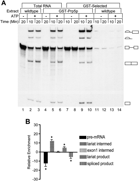
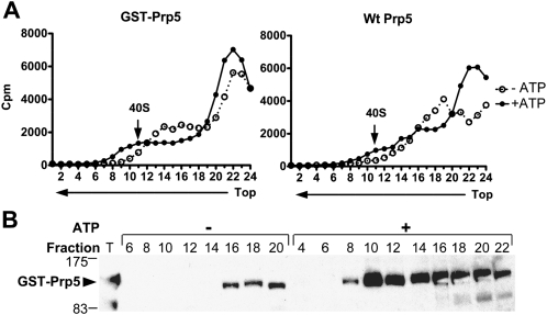
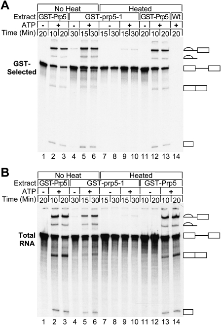
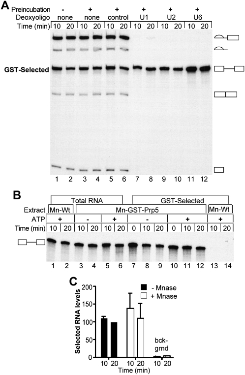
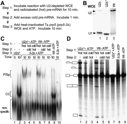
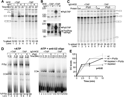
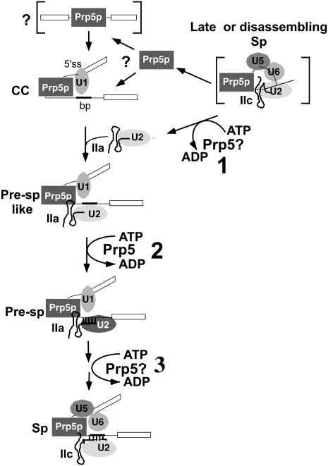
The DExD/H-box Prp5 protein (Prp5p) is an essential, RNA-dependent ATPase required for pre-spliceosome formation during nuclear pre-mRNA splicing. In order to understand how this protein functions, we used in vitro, biochemical assays to examine its association with the spliceosome from Saccharomyces cerevisiae. GST-Prp5p in splicing assays pulls down radiolabeled pre-mRNA as well as splicing intermediates and lariat product, but reduced amounts of spliced mRNA. It cosediments with active spliceosomes isolated by glycerol gradient centrifugation. In ATP-depleted extracts, GST-Prp5p associates with pre-mRNA even in the absence of spliceosomal snRNAs. Maximal selection in either the presence or absence of ATP requires a pre-mRNA with a functional intron. Prp5p is present in the commitment complex and functions in subsequent pre-spliceosome formation. Reduced Prp5p levels decrease levels of commitment, pre-spliceosomal and spliceosomal complexes. Thus Prp5p is most likely an integral component of the spliceosome, being among the first splicing factors associating with pre-mRNA and remaining until spliceosome disassembly. The results suggest a model in which Prp5p recruits the U2 snRNP to pre-mRNA in the commitment complex and then hydrolyzes ATP to promote stable association of U2 in the pre-spliceosome. They also suggest that Prp5p could have multiple ATP-independent and ATP-dependent functions at several stages of the splicing cycle.


Figures

FIGURE 1.
FIGURE 1.
GST-Prp5p pulls down pre-mRNA, splicing intermediates, and lariat product from splicing reactions. (A) Total and selected RNAs from splicing reactions. Active whole-cell splicing extracts (WCEs) with wild-type Prp5p (lanes 1–3,11–14) or GST-Prp5p (lanes 4–10), were incubated at 23°C in splicing buffer with either glucose to deplete ATP or water. Radiolabeled actin pre-mRNA was then added with either no ATP (lanes 1,4,7,8,11,12) or ATP (lanes 2,3,5,6,9,10,13,14). Aliquots of splicing reactions were removed at the times indicated and incubated with glutathione-sepharose beads for 30 min at 4°C (lanes 7–14) or processed to isolate total RNA (lanes 1–6). One-tenth of total RNA and all of selected RNA samples were fractionated by denaturing gel electrophoresis and visualized by autoradiography. Lariat intermediate, lariat product, pre-mRNA, mature mRNA, and free exon 1 are indicated from the top to the bottom of the gel. (B) Relative RNA enrichment. Relative enrichment was calculated by subtracting the percentages of pre-mRNA, splicing intermediates, and products in unselected (total) isolated RNA in each 10 min and 20 min sample from the values for paired GST-selected RNAs. Data for the 10 min and 20 min samples were then combined. The means (±SD) for five independent experiments are plotted with asterisks indicating that those means differ significantly from 0; all p ≤ 0.02.
FIGURE 2.
FIGURE 2.
Cosedimentation of GST-Prp5p with 40S spliceosomes in glycerol gradients. Radiolabeled AC/AC mutant pre-mRNA was incubated for 15 min in splicing reactions depleted of or supplemented with ATP, and with WCEs with either the N-terminal GST-tag, or untagged Prp5p. Reactions were then fractionated by sedimentation in 11%–23% glycerol gradients in low ionic conditions (50 mM KCl, 2 mM MgCl2, and 20 mM HEPES at pH 7.6). Gradient fractions were collected and analyzed. (A) Radioactive counts in each fraction. Cherenkov emissions in aliquots of gradient fractions from reactions with GST-Prp5p (left panel) or with untagged Prp5p (right panel) were counted. An arrow indicates the 40S spliceosomal peak (fraction 11). (Left panel, fractions 13–17 with GST-Prp5p) A shoulder in the radioactivity profile from reactions depleted of ATP occasionally occurred, but it did not depend on the presence of a tag. (B) Western blot of 40S fractions probed with anti-Prp5p antibody. Proteins in even-numbered fractions with GST-Prp5p with or without ATP (left panel in A) were concentrated by precipitation and analyzed by a Western blot probed with anti-Prp5p antibody. An aliquot of WCE with GST-Prp5p (T) corresponds to 20% of the reaction loaded onto the gradient. The bulk of GST-Prp5p in the gradient from the ATP-depleted reaction resides in fractions 22–24 (not shown). Molecular weight markers are indicated on the left. (Arrow) Indicates full-length GST-Prp5p. A fragment of GST-Prp5p (at 83 kDa) was probably generated by proteolytic cleavage during WCE preparation.
FIGURE 3.
FIGURE 3.
Heat-inactivated Ts GST-prp5-1 protein pulls down pre-mRNA from splicing reactions. (A) RNA in GST-selected material. Active WCE with GST-prp5-1p, GST-Prp5p, or wild-type Prp5p was either heated for 10 min at 37°C or incubated on ice (no heat) before adding splicing buffer with either glucose or water. After incubation for an additional 15 min at 30°C to deplete ATP, radiolabeled pre-mRNA and either no ATP (to the glucose treated reactions) or ATP (to water-treated reactions) was then added. Samples were removed at the times indicated and subjected to GST selection. RNA was extracted, fractionated by denaturing PAGE, and visualized by autoradiography (23 h exposure). (B) Total RNA from splicing reactions. RNA was extracted from samples removed from the reactions in A before GST selection. An amount from a sample equivalent to that in A was fractionated by denaturing PAGE and visualized by autoradiography (6 h exposure).
FIGURE 4.
FIGURE 4.
Efficient association of Prp5p with RNA requires a functional intron. (A) Diagram of actin wild-type and mutant pre-mRNAs. (From left to right) Bold lettering indicates the 5′ splice site, the cryptic branch site, the authentic branch site, and the 3′ splice site. Also depicted are mutant pre-mRNAs and the 6-nt cryptic branchpoint sequence deleted from the Δ6 transcript. The A1, A257, and AC/AC mutations were each made in the Δ6 background. (B) RNA in GST-Prp5p selected material. ATP-depleted GST-Prp5p (GST) or wild-type (WT) extracts in splicing buffer were incubated with equal amounts of functional actin (Δ6), 5′ splice site mutant (A1), branch site mutant (A257), and antisense (AS) RNAs for the indicated times. Splicing reactions were subjected to GST selection after which RNA was extracted, fractioned by denaturing PAGE, and visualized by autoradiography (12-h film exposure for lanes 1–10 and 96-h PhosphorImager screen exposure for lanes 11–18). (C) Total RNA from splicing reactions. RNA was extracted from samples removed from reactions before GST selection. RNA was visualized by autoradiography (2.5-h film exposure for lanes 1–12 and 67-h Phos-phorImager screen exposure for lanes 11–18). (D) Quantification of GST-selected RNA levels. Levels of selected wild-type and mutant A1 and A257 RNAs in three experiments as in A were measured and calculated as percentage of wild-type RNA pulled down at 20 min in the same experiment. The means (±SD) are plotted. Levels statistically significantly different from Δ6 levels are indicated by asterisks; all p ≤ 0.01. The levels of A1 and A257 are not statistically different from each other. Antisense RNA levels are the same as background levels (selections with an untagged Prp5 protein and selections with GST-tag only) measured in two, 10, and two experiments, respectively.
FIGURE 5.
FIGURE 5.
Association of Prp5p with pre-mRNA does not require intact U1, U2, or U6 RNA. (A) Pre-mRNA in GST-Prp5p selected material from reactions with RNase-H-degraded RNAs. Deoxyoligonucleotides complementary to the 5′ end regions of U1, U2, and U6 RNAs specifically target these RNAs for cleavage by endogenous RNase H activity. Active GST-Prp5p WCEs supplemented with 0.2 mM ATP were first incubated with added water or the following deoxyoligonucleotides: 1.0 μM control, 5.0 μM anti-U1, 1.0 μM anti-U2, and 1.0 μM of anti-U6 for 30 min at 30°C. The splicing reactions were initiated by adding radiolabeled pre-mRNA and fresh ATP and incubated at 23°C. Samples were removed at 10 and 20 min and subjected to GST-selection before RNA extraction and denaturing PAGE. GST-Prp5p WCE that did not undergo the first incubation at 30°C was used as an additional control (lanes 1,2). (B) GST-selected pre-mRNAs from micrococcal nuclease-treated WCE. WCEs of GST-Prp5p or wild-type Prp5p were first incubated with micrococcal nuclease (MNase) to destroy RNAs, after which reactions were stopped by adding EGTA. Splicing buffer with either glucose (−ATP) or water (+ATP) was then added to MNase-treated extracts (Mn-Wt or Mn-GST-Prp5p) and incubated for an additional 15 min at 30°C. After addition of fresh ATP (+ATP), radiolabeled pre-mRNA was added, and the reactions were incubated at 23°C. Samples were removed at the times indicated and either subjected to GST-selection (lanes 7–14) or processed to isolate total RNA (lanes 1–6). Zero time points were collected as described in Materials and Methods. Equal volumes of undiluted GST-selected material and 10-fold diluted total extracted RNA were fractionated by denaturing PAGE and visualized by autoradiography. (C) Quantification of RNAs selected from MNase-treated WCE. WCE with GST-Prp5p or untagged Prp5p (“bckgrnd”) was incubated with MNase (Mn-GST-Prp5p) or water and depleted of ATP. The extracts were then analyzed by pull-down assays as in B. Levels of selected RNAs in two experiments were measured and calculated as a percentage of RNA pulled down from reactions with untreated WCE at 20 min in the same experiment. The means (±SD) are plotted. There is no significant difference due to MNase treatment.
FIGURE 6.
FIGURE 6.
Prp5p in the commitment complex functions in pre-spliceosome formation. (A) Diagram of commitment-chase experiment. In Step 1, radiolabeled (hot), pre-mRNA was incubated for 10 min at 23°C in reactions with U2-depleted WCE to form commitment complexes. In Step 2, 190-fold excess unlabeled (cold) act pre-mRNA was added, and the reaction was incubated for 1 min. In Step 3, heat-inactivated Ts prp5-3 mutant WCE and ATP were added, and the reaction was incubated for 10 min. After Step 3, part of the reaction was analyzed by native gel electrophoresis, and the other part was analyzed for splicing activity. (B) Northern analysis of U2 RNA in WCEs used in the commitment experiment. RNA was extracted from equal amounts of wild-type WCE (Wt) and WCE from yeast cells metabolically depleted of U2 (U2Δ). U2Δ WCE was treated further by deoxyoligonucleotide-directed RNase H cleavage (U2Δ*) to inactivate any residual U2 before RNAs were extracted. RNAs were measured by Northern blot analysis with U1 and U2 RNA probes. (Solid arrowheads) Intact U2 and U1 RNAs. (Open arrowhead) U2 RNA degraded by RNase H cleavage. RNAse H treatment reduced levels of intact U2 to ≤ 2% that of wild type. (C) Spliceosomal complexes assayed by native gel electrophoresis. In Step 1, hot (lanes 3,4,6,7) or excess cold (lanes 5,8) pre-mRNA was first incubated in reactions with WCEs with wild-type Prp5p. Either U2-depleted (U2Δ*, lanes 3–5) or ATP-depleted wild type (Wt, lanes 6–8) WCE was used. In Step 2, water (lanes 3,6), excess cold pre-mRNA (lanes 4,7), or hot pre-mRNA (lanes 5,8) was added and the reactions were incubated for 1 min for Step 2. In Step 3, ATP and heat-inactivated prp5-3 WCE were added and the reactions were incubated for 10 min (lanes 38). Three control reactions were incubated with hot pre-mRNA for 10 min as follows: (lane 2) U2-depleted WCE (U2Δ*); (lane 9) ATP-depleted wild-type WCE; (lane 10) heat-inactivated prp5-3 WCE and ATP. Lane 1 is a “zero” time control in which U2Δ* WCE and splicing buffer are incubated with loading buffer and competitor RNA for 10 min on ice before hot pre-mRNA is added. In this native gel electrophoretic assay, the pre-spliceosomal and spliceosomal complexes comigrate, and CC1 and CC2 comigrate (Ruby 1997). Low levels of CC in the reaction with heat-inactivated prp5-3 WCE (lane 10) did not result from the prp5-3 mutation or interfere with the assay. (D) Splicing assay. In Step 1, hot (lanes 1,2,4,5) or excess cold (lanes 3,6) pre-mRNA was incubated in reactions with WCEs with wild-type Prp5p. Either U2-depleted (U2Δ*, lanes 1–3) or ATP-depleted wild type (Wt, lanes 4–6) WCE was used. In Step 2, water (lanes 1,4), excess cold pre-mRNA (lanes 2,5), or hot pre-mRNA (lanes 3,6) was added and the reactions were incubated for 1 min for Step 2. In Step 3, ATP and heat-inactivated prp5-3 WCE were added and the reactions were incubated for 10 min (lanes 1–6). Control reactions were incubated with hot pre-mRNA for 10 min as follows: (lane 7) U2-depleted WCE and ATP (U2Δ* + ATP); (lane 8) heat-inactivated prp5-3 WCE, and ATP; (lane 9) ATP-depleted wild-type WCE. Pre-mRNA and splicing intermediates and products are indicated as in Figure 1.
FIGURE 7.
FIGURE 7.
Decreased Prp5p levels reduce levels of splicing and spliceosomal complexes. (A) Splicing assay of depleted and mock-treated WCEs. WCEs with either TAP-tagged or untagged Prp5p were treated with IgG-Sepharose or mock-treated with unmodified Sepharose. The WCEs were then assayed for splicing activity using radiolabeled wild-type pre-mRNA. The spliced intermediates and products derived from the wild-type act pre-mRNA are depicted as in Figure 1A. The mean percent total spliced RNA from two assays was calculated as described in Materials and Methods and indicated at the bottom of each lane; standard deviations were <10% of the means. (B) Western analysis of depleted and mock-treated WCEs. Samples of WCEs with either untagged or TAP-tagged Prp5 protein and either mock-treated or treated with IgG-Sepharose were fractionated by SDS-PAGE. Prp5 protein was detected by Western blotting and by probing first with anti-CBP antibody to detect the TAP-tag (top panel) and second with anti-Prp5p antibody to detect the Prp5 protein (bottom panel). (Open arrowhead) Wild-type Prp5p. (Solid arrowheads) Prp5-TAP or prp5p fragments derived from Prp5-TAP (Prp5frag). Positions of molecular weight markers are indicated on the left. The blot was not stripped between probings, so the Prp5-TAP signal is stronger than the signals for the wild-type Prp5p due to residual anti-CBP antibody. (C) Complementation of depleted and mock-treated extracts. Recombinant GST (1.6 μM protein) or Prp5p (from 126 nM to 2 μM protein in twofold increments) was added to splicing reactions containing radiolabeled pre-mRNA and IgG-treated WCE (IgG-WCE) with originally either TAP-tagged or untagged Prp5p and incubated for 10 min at 23°C. Total RNA was extracted, fractionated by denaturing PAGE, and visualized with a PhosphorImager. The spliced intermediates and products derived from the wild-type act pre-mRNA are depicted as in Figure 1. The mean percent total spliced RNA from two (lanes 1–6) and three (lanes 7–12) assays was calculated as described in Materials and Methods, and are indicated at the bottom of each lane; standard deviations were <14% (lanes 1–6) and 23% (lanes 7–12) of the means. (D) Native gel electrophoretic assays of spliceosomal complexes in depleted and mock-treated extracts. Radiolabeled wild-type act pre-mRNA was added to splicing reactions with IgG-treated WCEs (IgG-WCE) with either Prp5-TAP or untagged Prp5p and with or without added recombinant wild-type Prp5p. Samples were removed at the times indicated and fractionated by native gel electrophoresis. The complexes were visualized and measured with a PhosphorImager. The pre-spliceosomal and spliceosomal complexes migrate close to one another and appear as one band (p/sp). Only one CC band is formed in this native gel electrophoretic assay. (Left panel) ATP was added to the splicing reactions. (Bottom of each lane) The percentage of RNA in p/sp [p/sp divided by (CC + p/sp)] is listed. (Right panel) The WCEs were incubated with glucose to deplete ATP and a 2′-O-methyl oligonucleotide complementary to U2 RNA to block pre-spliceosome formation before radiolabeled pre-mRNA was added. (E) Quantification of CC levels. CC levels as measured in experiments in the right panel of D were calculated as the percentage of the maximal levels reached in that particular extract supplemented with rPrp5p. The means (±SD) were calculated from three and two experiments for IgG-treated WCEs with either TAP-tag or untagged Prp5p, respectively.
FIGURE 8.
FIGURE 8.
Model of Prp5p in the splicing cycle. In this working model, the DEAD-box Prp5 protein in a late or disassembling spliceosome hydrolyzes ATP to convert U2 RNA from the IIC form to the IIA form (“1” in upper right corner). The bracketed complex indicates that the conversion of U2 could occur anytime after exon ligation through disassembly of the spliceosome. (Left side of diagram) U2 and Prp5 are recycled for a new round of splicing. The bracketed complex with pre-mRNA and Prp5p may precede CC, but the order of Prp5p and U1 snRNP binding to pre-mRNA is uncertain. In the commitment complex (CC), Prp5p may interact with U1 snRNP and other proteins to bridge the 5′ splice site and the UACUAACA box. It may also recruit U2 snRNP via protein–protein interactions in the absence of ATP to form a pre-spliceosome-like complex (Pre-Sp-like) in which U2 RNA is not yet paired with pre-mRNA. Prp5p then hydrolyzes ATP (“2” in the diagram) to form the stable pre-spliceosome (Pre-Sp) with U2 base-paired with pre-mRNA. Finally, Prp5p may have additional ATP-dependent functions after pre-spliceosome formation (“3” in the diagram) and switch U2 between the IIA and IIC forms. Only those splicing factors relevant to this study are shown, and they are arbitrarily arranged relative to one another. Prp5p may or may not bind pre-mRNA directly.

References

    1. Abovich N, Rosbash M. Cross-intron bridging interactions in the yeast commitment complex are conserved in mammals. Cell. 1997;89:403–412. - PubMed
    1. Abu Dayyeh B, Quan TK, Castro MA, Ruby SW. Probing interactions between the U2 snRNP and the DEAD-box protein, Prp5. J Biol Chem. 2002;277:20221–20233. - PubMed
    1. Berglund JA, Chua K, Abovich N, Reed R, Rosbash M. The splicing factor BBP interacts specifically with the pre-mRNA branchpoint sequence UACUAAC. Cell. 1997;89:781–787. - PubMed
    1. Berglund N, Colot H, Rosbash M. A cooperative interaction between U2AF65 and mBBP/SF1 facilitates branchpoint region recognition. Genes & Dev. 1998;12:858–867. - PMC - PubMed
    1. Brody E, Abelson J. The “spliceosome”: Yeast pre-messenger RNA associates with a 40S complex in a splicing-dependent reaction. Science. 1985;228:963–967. - PubMed

Publication types

MeSH terms