. 2009 Jul 16;114(3):580-8.
doi: 10.1182/blood-2009-01-200923. Epub 2009 May 18.

CCL21 mediates CD4+ T-cell costimulation via a DOCK2/Rac-dependent pathway

Affiliations

CCL21 mediates CD4+ T-cell costimulation via a DOCK2/Rac-dependent pathway

Kathrin Gollmer et al. Blood. .

Abstract

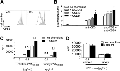
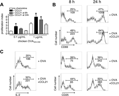
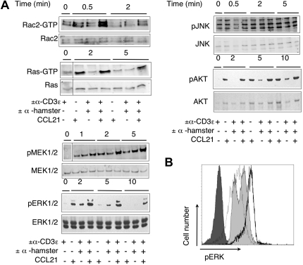
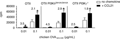
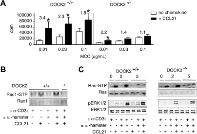
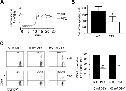
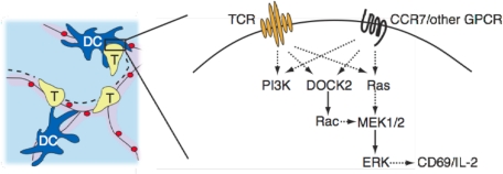
CD4(+) T cells use the chemokine receptor CCR7 to home to and migrate within lymphoid tissue, where T-cell activation takes place. Using primary T-cell receptor (TCR)-transgenic (tg) CD4(+) T cells, we explored the effect of CCR7 ligands, in particular CCL21, on T-cell activation. We found that the presence of CCL21 during early time points strongly increased in vitro T-cell proliferation after TCR stimulation, correlating with increased expression of early activation markers. CCL21 costimulation resulted in increased Ras- and Rac-GTP formation and enhanced phosphorylation of Akt, MEK, and ERK but not p38 or JNK. Kinase-dead PI3Kdelta(D910A/D910A) or PI3Kgamma-deficient TCR-tg CD4(+) T cells showed similar responsiveness to CCL21 costimulation as control CD4(+) T cells. Conversely, deficiency in the Rac guanine exchange factor DOCK2 significantly impaired CCL21-mediated costimulation in TCR-tg CD4(+) T cells, concomitant with impaired Rac- but not Ras-GTP formation. Using lymph node slices for live monitoring of T-cell behavior and activation, we found that G protein-coupled receptor signaling was required for early CD69 expression but not for Ca(2+) signaling. Our data suggest that the presence of CCL21 during early TCR signaling lowers the activation threshold through Ras- and Rac-dependent pathways leading to increased ERK phosphorylation.


Figures

Figure 1
Figure 1
Homeostatic chemokines function as costimulatory factors during Ab- and peptide-induced T-cell activation. (A) Flow cytometry histogram of CFSE-labeled T-cell division after 48 and 72 hours of activation with anti-CD3ϵ mAb in the presence (bold line) or absence (gray fill) of 100 nM CCL21. Data are representative from 1 of 6 independent experiments. (B) Proliferation in the presence and absence of homeostatic chemokines. CFSE-labeled T cells were stimulated with plate-bound anti-CD3ϵ mAb with or without anti-CD28 mAb in the presence or absence of CCL21, CCL19, or CXCL12 (100 nM). Proliferation was determined after 48 hours by fluorescence-activated cell sorter and normalized to fold increase compared with cells cultured with anti-CD3ϵ mAb (proliferation index). The presence of chemokines increases the percentage of cells having undergone cell divisions within 48 hours. Data are pooled from 4 to 6 independent experiments. Statistical significance was determined using analysis of variance comparison of anti-CD3 or anti-CD3/CD28–stimulated T-cell proliferation with or without chemokines. *P < .05 compared with “no chemokine.” (C) DO11.10 T cells were cocultured with chicken or turkey OVA323-339-loaded congenic splenocytes in the presence or absence of CCL21 (100 nM). T-cell activation was determined by 3H-thymidine-incorporation after 48 hours. Numbers indicate fold increase of proliferation in the presence of CCL21. *P < .05 compared with “no chemokine.” (D) Prior activation as in panel C; DO11.10 T cells were incubated with PTX. Data are pooled from 2 independent experiments.
Figure 2
Figure 2
CCL21 costimulation is effective during early T-cell activation. DO11.10 T cells were cocultured with OVA323-339-loaded congenic splenocytes in the presence or absence of CCL21 (100 nM). (A) CCL21 was added at indicated times, and proliferation of T cells was analyzed after 48 hours. Statistical significance was determined using analysis of variance comparison of peptide-stimulated T-cell proliferation without or with CCL21 added at indicated times. *P < .05 compared with “no chemokine.” (B) Up-regulation of early activation markers CD69 and CD25 on CD4+ KJ1-26+ DO11.10 T cells 8 and 24 hours after Ag-specific T-cell activation in the presence or absence of CCL21 (100 nM). One representative experiment of 3 is shown. (C) IL-2 production of DO11.10 T cells after 24 hours as determined by intracellular staining. One representative experiment of 2 is shown. In panels B and C, numbers indicate percentage of positive cells and mean fluorescence intensity (MFI), respectively.
Figure 3
Figure 3
Biochemical analysis of CCL21 costimulation. Primary mouse T cells were stimulated with anti-CD3 crosslinking, in the presence or absence of CCL21 (100 nM), for indicated times, and the activation of early signaling molecules was analyzed by immunoblotting. (A) Immunoblots of Rac2-GTP, Ras-GTP, and phosphorylated MEK1/2, ERK1/2, JNK, and Akt after costimulation of TCR and/or CCL21. For loading controls, blots were stripped and probed for total protein, or alternatively, a separate gel with lysates was analyzed. (B) Flow cytometric analysis of phosphorylated ERK1/2 formation in nonstimulated primary mouse T cells (dark gray fill) or after 5 minutes of stimulation with CCL21 (100 nM; dashed line), TCR-crosslinking (light gray fill), and TCR-crosslinking in the presence of 100 nM CCL21 (bold line). One representative experiment of 2 is shown.
Figure 4
Figure 4
CCL21-mediated costimulation in the absence of PI3Kγ- or PI3Kδ-activity. OT-II TCR-tg CD4+ T cells were cocultured with chicken OVA323-339-pulsed irradiated congenic splenocytes in the presence or absence of CCL21 (100 nM). T-cell activation was determined as described in “Proliferation assays.” Proliferation of control, PI3Kγ-deficient, and PI3KδD910A/D910A OT-II TCR-tg CD4+ T cells after 72 hours is shown. Numbers indicate fold increase of proliferation in the presence of CCL21. Data are pooled from 4 independent experiments. *P < .05 compared with “no chemokine.”
Figure 5
Figure 5
CCL21-mediated costimulation and signaling pathways in DOCK2-deficient T cells. (A) 2B4 TCR-tg CD4+ T cells were cocultured with MCC88-103-pulsed irradiated splenocytes in the presence or absence of CCL21 (100 nM). T-cell activation was determined as described in “Proliferation assays.” Proliferation of TCR-tg DOCK2+/− and DOCK2−/− 2B4 TCR-tg T cells after 48 hours is shown. Numbers indicate fold increase of proliferation in the presence of CCL21. Data are pooled from 3 independent experiments. *P < .05 compared with “no chemokine.” (B) Control and DOCK2−/− 2B4 TCR-tg T cells were stimulated as in Figure 3 and analyzed for Rac-GTP formation. One representative experiment of 2 is shown. (C) Control and DOCK2−/− 2B4 TCR-tg T cells were stimulated as in Figure 3 and analyzed for phosphorylation of ERK1/2 and Ras-GTP formation. One representative experiment of 3 is shown.
Figure 6
Figure 6
Antigen-induced T-cell responses measured in PLN slices are decreased by PTX. (A) Average Ca2+ responses of Marylin TCR-tg CD4+ T cells incubated with suB or PTX and stimulated with DBY peptide. Fura-2-loaded T cells were incubated for 10 minutes with 100 ng/mL suB (black line) or PTX (gray line), washed, and overlaid on PLN slices. Time-lapse imaging was started 2 hours after suB or PTX treatment. After several minutes of imaging, the preparation was perfused with a solution containing 100 nM DBY peptide. Ca2+-signals of responding T cells were synchronized at the rising phases of the response. (B) Percentage of Ca2+-responding CD4+ T cells induced by DBY peptide (100 nM). Mean plus or minus SD of 3 independent experiments in which more than 50 cells were analyzed per experiment. *P < .05. (C) CD69 expression measured by flow cytometry on T cells activated within PLN slices. Marylin CD4+ TCR-tg T cells were incubated with 100 ng/mL of suB or PTX, labeled with CMFDA and overlaid on PLN slices. Two hours after DBY peptide treatment, slices were mechanically dissociated and recovered cells stained with anti–CD69-PE and anti–CD45.1-PE-Cy5 Abs. Results in the left panel are representative of 3 independent experiments and show percentage and MFI of CD69+ cells. The right panel shows combined MFI of CD69 expression after normalization to the value of suB-treated T cells. *P < .05.
Figure 7
Figure 7
Proposed model for integration of TCR and CCR7-triggered signals. During an early promigratory phase of an ongoing antigenic response, CD4+ T cells undergo sequential encounters with pMHC-presenting DCs (left panel). During this period, T cells are exposed to TCR- and CCR7 (in addition to other GPCR)-derived signals, both of which activate PI3K, DOCK2-Rac, and Ras. Active Rac- and Ras-GTP contribute to enhanced MEK-ERK activation leading to up-regulation of early activation markers, such as CD69, and rapid production of IL-2 (right panel).

References

    1. von Andrian UH, Mempel TR. Homing and cellular traffic in lymph nodes. Nat Rev Immunol. 2003;3:867–878. - PubMed
    1. Lanzavecchia A, Sallusto F. Dynamics of T lymphocyte responses: intermediates, effectors, and memory cells. Science. 2000;290:92–97. - PubMed
    1. Stoll S, Delon J, Brotz TM, Germain RN. Dynamic imaging of T cell-dendritic cell interactions in lymph nodes. Science. 2002;296:1873–1876. - PubMed
    1. Mempel TR, Henrickson SE, Von Andrian UH. T-cell priming by dendritic cells in lymph nodes occurs in three distinct phases. Nature. 2004;427:154–159. - PubMed
    1. Miller MJ, Wei SH, Parker I, Cahalan MD. Two-photon imaging of lymphocyte motility and antigen response in intact lymph node. Science. 2002;296:1869–1873. - PubMed

Publication types