Developmental sources of conservation and variation in the evolution of the primate eye
- PMID: 19451636
- PMCID: PMC2690025
- DOI: 10.1073/pnas.0901484106
Developmental sources of conservation and variation in the evolution of the primate eye
Abstract
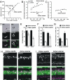
Conserved developmental programs, such as the order of neurogenesis in the mammalian eye, suggest the presence of useful features for evolutionary stability and variability. The owl monkey, Aotus azarae, has developed a fully nocturnal retina in recent evolution. Description and quantification of cell cycle kinetics show that embryonic cytogenesis is extended in Aotus compared with the diurnal New World monkey Cebus apella. Combined with the conserved mammalian pattern of retinal cell specification, this single change in retinal progenitor cell proliferation can produce the multiple alterations of the nocturnal retina, including coordinated reduction in cone and ganglion cell numbers, increase in rod and rod bipolar numbers, and potentially loss of the fovea.
Conflict of interest statement
The authors declare no conflict of interest.
Figures


Similar articles
-
Horizontal cell morphology in nocturnal and diurnal primates: a comparison between owl-monkey (Aotus) and capuchin monkey (Cebus).Vis Neurosci. 2005 Jul-Aug;22(4):405-15. doi: 10.1017/S0952523805224033. Vis Neurosci. 2005. PMID: 16212699
-
Distribution of M retinal ganglion cells in diurnal and nocturnal New World monkeys.J Comp Neurol. 1996 May 13;368(4):538-52. doi: 10.1002/(SICI)1096-9861(19960513)368:4<538::AID-CNE6>3.0.CO;2-5. J Comp Neurol. 1996. PMID: 8744442
-
The Heterochromatin Block That Functions as a Rod Cell Microlens in Owl Monkeys Formed within a 15-Myr Time Span.Genome Biol Evol. 2021 Mar 1;13(3):evab021. doi: 10.1093/gbe/evab021. Genome Biol Evol. 2021. PMID: 33533923 Free PMC article.
-
Morphology and physiology of primate M- and P-cells.Prog Brain Res. 2004;144:21-46. doi: 10.1016/S0079-6123(03)14402-0. Prog Brain Res. 2004. PMID: 14650838 Review.
-
Developmental duration as an organizer of the evolving mammalian brain: scaling, adaptations, and exceptions.Evol Dev. 2020 Jan;22(1-2):181-195. doi: 10.1111/ede.12329. Epub 2019 Dec 3. Evol Dev. 2020. PMID: 31794147 Review.
Cited by
-
The AII amacrine cell connectome: a dense network hub.Front Neural Circuits. 2014 Sep 4;8:104. doi: 10.3389/fncir.2014.00104. eCollection 2014. Front Neural Circuits. 2014. PMID: 25237297 Free PMC article.
-
Evo-devo and brain scaling: candidate developmental mechanisms for variation and constancy in vertebrate brain evolution.Brain Behav Evol. 2011;78(3):248-57. doi: 10.1159/000329851. Epub 2011 Aug 23. Brain Behav Evol. 2011. PMID: 21860220 Free PMC article. Review.
-
The Dynamic Epigenetic Landscape of the Retina During Development, Reprogramming, and Tumorigenesis.Neuron. 2017 May 3;94(3):550-568.e10. doi: 10.1016/j.neuron.2017.04.022. Neuron. 2017. PMID: 28472656 Free PMC article.
-
MicroRNA Signatures of the Developing Primate Fovea.Front Cell Dev Biol. 2021 Apr 8;9:654385. doi: 10.3389/fcell.2021.654385. eCollection 2021. Front Cell Dev Biol. 2021. PMID: 33898453 Free PMC article.
-
Genetic Dissection of Dual Roles for the Transcription Factor six7 in Photoreceptor Development and Patterning in Zebrafish.PLoS Genet. 2016 Apr 8;12(4):e1005968. doi: 10.1371/journal.pgen.1005968. eCollection 2016 Apr. PLoS Genet. 2016. PMID: 27058886 Free PMC article.
References
-
- Dawkins R. The Blind Watchmaker. New York: Norton; 1986.
-
- Land M, Nilsson D. Animal Eyes. Oxford: Oxford Univ Press; 2002.
-
- Fernald RD. Evolution of eyes. Curr Opin Neurobiol. 2000;10:444–450. - PubMed
-
- Kirschner MW, Gerhart JC. The Plausibility of Life: Resolving Darwin's Dilemma. New Haven, CT: Yale Univ Press; 2005.
-
- Teotonio H, Rose MR. Variation in the reversibility of evolution. Nature. 2000;408:463–466. - PubMed
Publication types
MeSH terms
LinkOut - more resources
Full Text Sources
Medical

