. 2009 Jul 31;284(31):20885-96.
doi: 10.1074/jbc.M109.016584. Epub 2009 May 18.

Farnesylation of pex19p is required for its structural integrity and function in peroxisome biogenesis

Affiliations

Farnesylation of pex19p is required for its structural integrity and function in peroxisome biogenesis

Robert Rucktäschel et al. J Biol Chem. .

Abstract

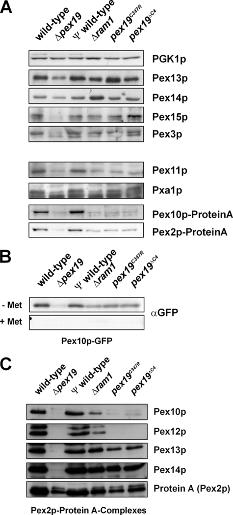
The conserved CaaX box peroxin Pex19p is known to be modified by farnesylation. The possible involvement of this lipid modification in peroxisome biogenesis, the degree to which Pex19p is farnesylated, and its molecular function are unknown or controversial. We resolve these issues by first showing that the complete pool of Pex19p is processed by farnesyltransferase in vivo and that this modification is independent of peroxisome induction or the Pex19p membrane anchor Pex3p. Furthermore, genomic mutations of PEX19 prove that farnesylation is essential for proper matrix protein import into peroxisomes, which is supposed to be caused indirectly by a defect in peroxisomal membrane protein (PMP) targeting or stability. This assumption is corroborated by the observation that mutants defective in Pex19p farnesylation are characterized by a significantly reduced steady-state concentration of prominent PMPs (Pex11p, Ant1p) but also of essential components of the peroxisomal import machinery, especially the RING peroxins, which were almost depleted from the importomer. In vivo and in vitro, PMP recognition is only efficient when Pex19p is farnesylated with affinities differing by a factor of 10 between the non-modified and wild-type forms of Pex19p. Farnesylation is likely to induce a conformational change in Pex19p. Thus, isoprenylation of Pex19p contributes to substrate membrane protein recognition for the topogenesis of PMPs, and our results highlight the importance of lipid modifications in protein-protein interactions.


Figures

FIGURE 1.
FIGURE 1.
Pex19p is completely farnesylated in vivo, independent of peroxisome induction and Pex3p. A and B, Pex19p is fully modified by yeast FTase in vivo. Whole cell lysates from non-induced cells of the indicated strains were analyzed by immunoblotting. Blots were probed with anti-Pex19p antibodies. The non-farnesylated form of Pex19p of a Δram1 mutant (arrowhead) cannot be detected in extracts from wild-type yeast (arrow) (A), whereas it reappears after reintroduction of Ram1p (B). C, the yeast farnesylation machinery can be saturated by overexpression of GST-Pex19p. A Coomassie-stained gel of purified farnesylated and non-farnesylated Pex19p is shown. GST-Pex19p was expressed under control of a copper-inducible promoter in Δpex19 and Δram1 strains and isolated by affinity chromatography. In Δram1 (right), only the non-farnesylated GST-Pex19p can be detected. In Δpex19 (left) two bands appear, corresponding to non-farnesylated GST-Pex19p (upper band) and farnesylated GST-Pex19p (lower band). D, Pex19p farnesylation levels are independent of peroxisome induction and are not affected by the absence of the Pex19p membrane anchor Pex3p. Cells were grown on YPD medium and, where indicated, washed and grown on 0.1% oleate medium for 17 h for peroxisome induction. Lysates were fractionated by centrifugation (20,000 × g, 1 h, 4 °C) and analyzed as in A. Blots were probed with antibodies against Pex19p. E, evolutionary conservation of the Pex19p farnesylation site in fungi, plant, and metazoa.
FIGURE 2.
FIGURE 2.
Pex19p farnesylation is required for peroxisome function. A, genomic Pex19p mutants. In all cases, the kanMX4 marker was used to select for integration into the genome. CKQQ, the terminal four amino acids of wild-type S. cerevisiae Pex19p (CaaX box). For generation of the pseudo-wild type (ψ wild-type), the kanMX4 marker was introduced after the STOP codon of wild-type PEX19. In the pex19C347R mutant, the cysteine of the farnesylation site was genomically replaced by arginine. In the pex19ΔC4 strain, the farnesylation site CKQQ was removed by inserting a STOP codon followed by kanMX4 after PEX19 base pair 1038, corresponding to amino acid 346. B, appearance of Pex19p modification in genomic pex19 farnesylation mutants. The indicated strains were grown on glucose and oleate medium and analyzed by immunoblot with the antibodies indicated. PGK1p was used as loading control. C, growth assay on oleate liquid medium. Strains were precultured in synthetic medium (SD) with 0.3% glucose, washed, and inoculated at 0.05 A600 units/ml in 0.1% oleate and 2% ethanol medium. At the indicated time points, 1-ml samples were taken, sedimented by centrifugation, and washed, and A600 was determined.
FIGURE 3.
FIGURE 3.
Matrix protein import is disturbed in farnesylation mutants. A, subcellular localization of the peroxisomal marker GFP-PTS1. Cells expressing peroxisomal targeting signal 1 (SKL) fused to GFP were grown on synthetic medium (SD) with 0.1% oleate for 3 days and analyzed by direct fluorescence microscopy. Bar, 5 μm. B, separation of cytosolic and organellar fractions by differential centrifugation. Cell-free postnuclear supernatants of the indicated strains were fractionated by centrifugation into a soluble supernatant and a particular pellet fraction. Equal amounts of obtained total (T), supernatant (S), and pellet (P) fractions were subjected to immunoblot analysis with antibodies raised against peroxisomal catalase (Cta1p) and oxo-acyl-CoA thiolase (Fox3p) or by catalase activity measurements (C). The total activity of Δpex19 was set as 100%.
FIGURE 4.
FIGURE 4.
Pex19p farnesylation mutants are defective in PMP stability and targeting. A, whole cell lysates of oleic acid-induced cells were analyzed by SDS-PAGE and immunoblotting. Blots were decorated with the indicated antibodies. Some PMPs show reduced steady-state levels due to the lack of Pex19p or Pex19p-farnesylation. B, the indicated strains expressing Pex10p-GFP under the control of the MET25 promoter were grown either in the absence or presence of methionine in the culture media. Δram1 and strains expressing mutated Pex19p variants exhibit reduced Pex10p-GFP levels upon induction of gene expression. C, Pex2p-Protein A was immunoprecipitated from digitonin-solubilized membranes of the indicated strains. Precipitates were analyzed by SDS-PAGE and immunoblotting with the indicated antibodies. Strains expressing Pex19p-farnesylated mutants exhibited a highly reduced association of Pex2p with the other peroxisomal RING finger proteins, Pex10p and Pex12p.
FIGURE 5.
FIGURE 5.
In vitro farnesylation and structural difference between Pex19p and Pex19pFARN. A, expression and in vitro farnesylation of Pex19p and Ras1p. Pex19p and Ras1p were expressed in E. coli as GST fusion proteins, farnesylated by purified recombinant FTase, and analyzed by SDS-PAGE and Coomassie staining. The farnesylated forms of both proteins show a higher mobility on SDS gels. B, farnesylation was confirmed by electron spray ionization mass spectrometry. Non-farnesylated (left) and farnesylated (right) Pex19p were analyzed by ESI-MS. The expected molecular mass of the tag-free non-farnesylated form of Pex19p is 39,937 Da (including linker amino acids Gly-Ser-His). The molecular mass of the farnesyl group is 204 Da, so the expected molecular weight of Pex19pFARN corresponds to 40,141 Da. C, size exclusion and anion exchange chromatography. GST-Pex19p was prepared as in A, cleaved by thrombin, and analyzed by gel filtration (Superdex 200 pg; GE Healthcare) or anion-exchange chromatography (ResourceQ; GE Healthcare). Solid line, Pex19p; dashed line, Pex19pFARN. AU, absorbance units. D, Pex19p was prepared as in C and treated with trypsin at 30 °C. Samples were taken at the indicated time points and analyzed by SDS-PAGE and Coomassie staining. The arrows indicate salient differences in the protein pattern. E, CD spectra of Pex19p and Pex19pFARN. F, increase in α-helical domains upon farnesylation. A conformational change was deduced from independent structural predictions of Pex19p and Pex19pFARN.
FIGURE 6.
FIGURE 6.
Pex19p farnesylation is required for efficient association with PMPs. A, Pex19p farnesylation enhances binding to its binding site in several PMPs. Overlapping pentadecameric peptides sampling the Pex19p binding site of several PMPs with two-amino acid shifts between neighboring peptides were spotted onto nitrocellulose membranes in duplicate and tested for Pex19p interaction. Membranes were probed with equal amounts of purified GST-Pex19p or GST-Pex19pFARN. Bound protein was detected by monoclonal anti-GST antibodies. The numbers denote the amino acid positions that delimit the analyzed binding site. Farnesylation of Pex19p increased the affinity to its binding site for all PMPs tested. Pex13p-(1–31) served as a negative control. B and C, two-hybrid analysis. AD/BD, GAL4 activation/binding domain; wt, PEX19 wild-type; mut, pex19C347R. B, Pex19p binding to PMPs was tested with the Pex13p loop domain (PEX13L, amino acids 173–258) as well as full-length PEX11 and ANT1. Inset, immunoblot of two-hybrid whole cell lysates decorated with anti-Pex19p antibodies. C, efficient two-hybrid interaction with Pex3p requires Pex19p farnesylation. D, quantification of Pex19p PMP peptide binding by fluorescence polarization titration of a fluorescein isothiocyanate-Pex13p peptide with Pex19p and Pex19pFARN. Calculation of binding constants revealed a KD of 64 and 7.6 nm for Pex19p and Pex19pFARN, respectively. AD, activation domain; BD, binding domain; wt, wild type; mut, mutant; FITC, fluorescein isothiocyanate.

Similar articles

Cited by

References

    1. Young S. G., Fong L. G., Michaelis S. (2005) J. Lipid Res. 46, 2531–2558 - PubMed
    1. Sebti S. M., Der C. J. (2003) Nat. Rev. Cancer 3, 945–951 - PubMed
    1. Zhang F. L., Casey P. J. (1996) Annu. Rev. Biochem. 65, 241–269 - PubMed
    1. Magee T., Seabra M. C. (2005) Curr. Opin. Cell Biol. 17, 190–196 - PubMed
    1. Sinensky M. (2000) Biochim. Biophys. Acta 1529, 203–209 - PubMed

Publication types

MeSH terms

LinkOut - more resources