. 2009 Jun;119(6):1571-82.
doi: 10.1172/JCI37480. Epub 2009 May 18.

Abrogation of TGF-beta signaling enhances chemokine production and correlates with prognosis in human breast cancer

Affiliations

Abrogation of TGF-beta signaling enhances chemokine production and correlates with prognosis in human breast cancer

Brian Bierie et al. J Clin Invest. 2009 Jun.

Abstract

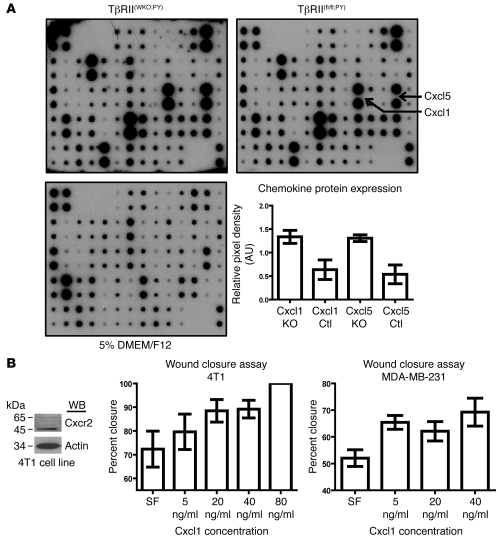
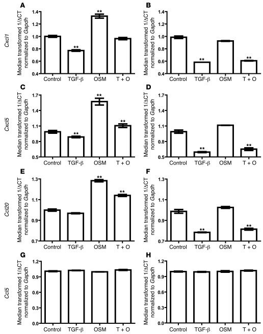
In human breast cancer, loss of carcinoma cell-specific response to TGF-beta signaling has been linked to poor patient prognosis. However, the mechanisms through which TGF-beta regulates these processes remain largely unknown. In an effort to address this issue, we have now identified gene expression signatures associated with the TGF-beta signaling pathway in human mammary carcinoma cells. The results strongly suggest that TGF-beta signaling mediates intrinsic, stromal-epithelial, and host-tumor interactions during breast cancer progression, at least in part, by regulating basal and oncostatin M-induced CXCL1, CXCL5, and CCL20 chemokine expression. To determine the clinical relevance of our results, we queried our TGF-beta-associated gene expression signatures in 4 human breast cancer data sets containing a total of 1,319 gene expression profiles and associated clinical outcome data. The signature representing complete abrogation of TGF-beta signaling correlated with reduced relapse-free survival in all patients; however, the strongest association was observed in patients with estrogen receptor-positive (ER-positive) tumors, specifically within the luminal A subtype. Together, the results suggest that assessment of TGF-beta signaling pathway status may further stratify the prognosis of ER-positive patients and provide novel therapeutic approaches in the management of breast cancer.


Figures

Figure 1
Figure 1. Recombination of TβRII and induction of TGF-β–dependent EMT in MMTV-PyVmT mammary carcinoma cells.
(A) Analysis of Southern blot hybridization demonstrated that the independently derived polyclonal TβRII(fl/fl;PY) control mammary carcinoma cells (FL1, FL2, and FL3; biological replicates) had intact floxed Tgfbr2 alleles, with no evidence of recombination. Alternatively, the independently derived polyclonal TβRII(WKO;PY) carcinoma cells (KO1, KO2, and KO3; biological replicates) were completely recombined, with no evidence of a remaining floxed Tgfbr2 allele. (B) The growth of TβRII(fl/fl;PY) control carcinoma cell lines, measured by tritiated thymidine incorporation 24 hours after stimulation, was markedly inhibited by TGF-β stimulation, whereas the TβRII(WKO;PY) carcinoma cell growth was not altered. Results represent median transformed mean values ± SEM. (C and D) In response to TGF-β stimulation (10 ng/ml) for 48 hours, the TβRII(fl/fl;PY) control carcinoma cells demonstrated consistent changes in morphology and cell scattering that suggested that EMT had occurred (original magnification, ×10). (EH) Loss of E-cadherin (E and F) and changes in F-actin localization from the cell membrane (G) to predominant association with stress fibers (H) further confirmed an EMT-like state in TβRII(fl/fl;PY) cells that had been treated with 10 ng/ml of TGF-β for 48 hours (EH; original magnification, ×40). (I and J) Loss of TGF-β signaling in TβRII(WKO;PY) cells did not result in a spontaneous state of EMT, as determined by the presence of membrane-bound E-cadherin, in the presence or absence of TGF-β ligand at 10 ng/ml after 48 hours of stimulation (original magnification, ×40).
Figure 2
Figure 2. TGF-β–dependent chemokine protein secretion by mammary carcinoma cells and the effect of Cxcl1 stimulation on metastatic mammary carcinoma cell migration.
(A) Conditioned medium from TβRII(WKO;PY) and TβRII(fl/fl;PY) mammary carcinoma cells revealed increased secretion of Cxcl1 and Cxcl5 protein by the TβRII(WKO;PY) populations according to cytokine antibody array. Quantitation of Cxcl1 and Cxcl5 expression was performed and represented as the median transformed mean values ± SEM. P < 0.05, TβRII(WKO;PY) (KO) vs. TβRII(fl/fl;PY) (Ctl), 2-tailed unpaired t test. (B) Wound closure assays were used to determine the effect of Cxcl1 presence on metastatic carcinoma cell migration. Cxcr2 protein production by the 4T1 cell line was observed by Western blot (WB), and the expression of CXCR2 by MDA-MD-231 cells has been previously reported (–40). Values are reported as mean percentage ± SEM. For the 4T1 carcinoma cell line, P = 0.5018 at 5 ng/ml, P = 0.0853 at 20 ng/ml, P = 0.0588 at 40 ng/ml, P < 0.005 at 80 ng/ml, 2-tailed unpaired t test. For the MDA-MB-231 cell line: P = 0.0019 at 5 ng/ml, P = 0.0411 at 20 ng/ml, and P = 0.0068 at 40 ng/ml, 2-tailed, unpaired t test.
Figure 3
Figure 3. TGF-β attenuated basal and OSM-induced expression of Cxcl1, Cxcl5, and Ccl20 in mammary epithelium.
TGF-β (10 ng/ml) and OSM (100 ng/ml) stimulation was performed for 1 hour in vitro. Real-time PCR was performed using HC11 (A, C, E, and G) and NMuMG (B, D, F, and H) cell lines. The median transformed 1/ΔCt values are reported as mean ± SEM. TGF-β significantly decreased Cxcl1 and Cxcl5 expression in HC11 and Cxcl1, Cxcl5, and Ccl20 in NMuMG cells (AF). In the OSM-responsive HC11 cell line, OSM significantly upregulated Cxcl1, Cxcl5, and Ccl20 expression (A, C, and E). TGF-β significantly attenuated the effect of OSM with regard to Cxcl1, Cxcl5, and Ccl20 expression in the HC11 cell line. The NMuMG cell line, which did not respond to OSM in growth response assays or analysis of phospho-Stat3 by Western blot, did not demonstrate chemokine regulation by OSM (B, D, and F). Ccl5 expression was not altered by TGF-β or OSM treatment in the HC11 or NMuMG cell lines (G and H, respectively). **P < 0.05. T + O, combined administration of TGF-β and OSM.
Figure 4
Figure 4. Loss of TGF-β signaling in mammary carcinoma cells resulted in a signature that correlated with increased risk of relapse during human breast cancer progression.
(A) The TGF-β signature correlation with breast cancer RFS in all patients. TβRII(WKO;PY) and TβRII(fl/fl;PY) mammary carcinoma gene expression signatures were compared with profiles from 1,319 human breast cancer tissues. The TβRII(WKO;PY) and TβRII(fl/fl;PY) plus TGF-β treatment signatures were also used to determine the correlation with RFS (left and right columns, respectively). The TβRII(WKO;PY) signature significantly correlated with decreased RFS. No significant difference in RFS was observed in correlation with the TGF-β treatment gene expression signature. (B) In human LN+ breast cancer patients, the TβRII(WKO;PY) signature correlated with reduced RFS (left), whereas the TGF-β treatment signature did not have a significant correlation (right). (C) In LN patients, no significant correlations were observed. Red, high correlation (r > 0); black, low correlation (r < 0). The “high versus low” model is based on transforming the TGF-β signature correlation into a dichotomous variable (high: r > 0; low: r < 0), and the “continuous” model uses the untransformed correlation as a continuous variable. Association of these groups with RFS was evaluated with the log-rank test.
Figure 5
Figure 5. Loss of TGF-β signaling in mammary carcinoma cells resulted in a signature that may further differentiate ER-positive tumors and risk of relapse in breast cancer patients.
Increased risk of poor RFS when human ER+ and luminal A breast cancer (A and B, respectively; left) was associated with a TGF-β signaling–deficient mammary carcinoma cell gene expression signature. The TGF-β treatment signature did not correlate with a difference in RFS (A and B, right). Although a trend toward reduced RFS was present when the TβRII(WKO;PY) signature was associated with hormone only–treated breast cancer (C), neither signature was statistically significant. Red, high correlation (r > 0); black, low correlation (r < 0). Association of these groups with RFS was evaluated with the log-rank test.

References

    1. Gobbi H., et al. Transforming growth factor-beta and breast cancer risk in women with mammary epithelial hyperplasia. J. Natl. Cancer Inst. 1999;91:2096–2101. doi: 10.1093/jnci/91.24.2096. - DOI - PubMed
    1. Gobbi H., et al. Loss of expression of transforming growth factor beta type II receptor correlates with high tumour grade in human breast in-situ and invasive carcinomas. Histopathology. 2000;36:168–177. doi: 10.1046/j.1365-2559.2000.00841.x. - DOI - PubMed
    1. Sun L., et al. Expression of transforming growth factor beta type II receptor leads to reduced malignancy in human breast cancer MCF-7 cells. J. Biol. Chem. 1994;269:26449–26455. - PubMed
    1. Shipitsin M., et al. Molecular definition of breast tumor heterogeneity. Cancer Cell. 2007;11:259–273. doi: 10.1016/j.ccr.2007.01.013. - DOI - PubMed
    1. Ziv E., Cauley J., Morin P.A., Saiz R., Browner W.S. Association between the T29-->C polymorphism in the transforming growth factor beta1 gene and breast cancer among elderly white women: The Study of Osteoporotic Fractures. JAMA. 2001;285:2859–2863. doi: 10.1001/jama.285.22.2859. - DOI - PubMed

Publication types

MeSH terms

Substances