

Computed tomographic angiography as an adjunct to digital subtraction angiography for the pre-operative assessment of cerebral aneurysms
- PMID: 19452029
- PMCID: PMC2682841
- DOI: 10.2174/1874205X00903010001
Computed tomographic angiography as an adjunct to digital subtraction angiography for the pre-operative assessment of cerebral aneurysms
Abstract
Objectives: Computerized tomographic angiography (CTA) has emerged as a valuable diagnostic tool for the management of patients with cerebrovascular disease. The use of CTA in lieu of, or as an adjunct to, conventional cerebral angiography in the management of cerebral aneurysms awaits further experience. In this study, we evaluated the role of CTA specifically for the pre-operative assessment and planning of cerebral aneurysm surgery.
Patients and methods: We reviewed the relevant neuroimaging of all patients treated at Dartmouth Hitchcock Medical Center between January, 2001 and December, 2004 with a diagnosis of cerebral aneurysm and diagnostic evaluation with both CTA and conventional digital subtraction angiography (DSA) using standard imaging protocols. 32 patients underwent both CTA and DSA during the study period for a total of 36 aneurysms. Images were independently re-assesed by two neurosurgeons for information valuable for pre-operative surgical planning.
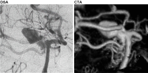
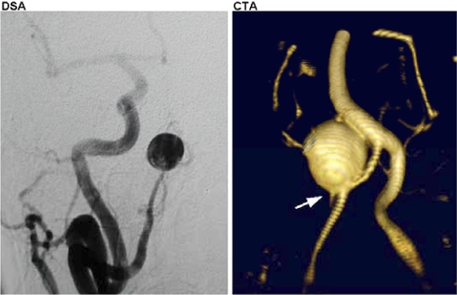
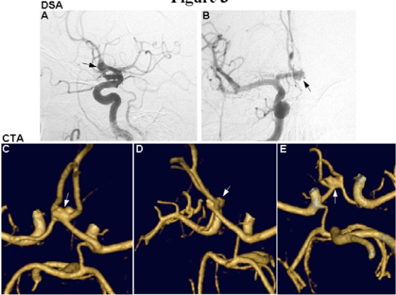
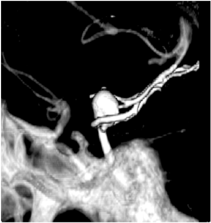
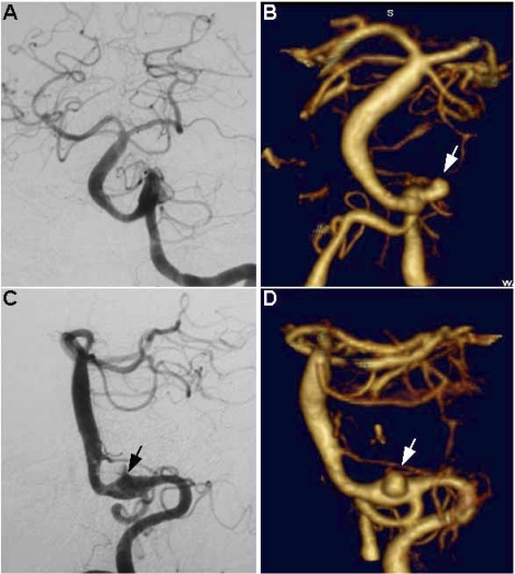
Results: In 26 of 36 aneurysms (72%), the CTA was felt to provide the best image quality in defining the morphology of the aneurysm. In 14 aneurysms (39%), CTA provided clinically valuable anatomic detail not demonstrated on DSA, largely due to better visualization of parent and perforating vessel relationships at the aneurysm neck. There were no instances where a lesion was seen on DSA but missed on CTA. The DSA was of most clinical value in determining flow dynamics, such as the arterial supply of an anterior communicating artery aneurysm and distal anterior cerebral branches via the two A1 segments.
Conclusion: CTA with three-dimensional reconstructions is a valuable adjunct to the preoperative evaluation of cerebral aneurysms. We advocate routine use of CTA in all patients in whom surgical aneurysm repair is planned, even when DSA has already been performed.
Keywords: Computed tomographic angiography; cerebral aneurysm; digital subtraction angiography; magnetic resonance angiography; pre-operative assessment..
Figures
Similar articles
-
Computed tomographic angiography versus digital subtraction angiography for the diagnosis and early treatment of ruptured intracranial aneurysms.Neurosurgery. 1999 Dec;45(6):1315-20; discussion 1320-2. doi: 10.1097/00006123-199912000-00008. Neurosurgery. 1999. PMID: 10598698
-
Experience with computed tomographic angiography for the detection of intracranial aneurysms in the setting of acute subarachnoid hemorrhage.Neurosurgery. 1997 Sep;41(3):522-7; discussion 527-8. doi: 10.1097/00006123-199709000-00003. Neurosurgery. 1997. PMID: 9310967
-
Results of a prospective protocol of computed tomographic angiography in place of catheter angiography as the only diagnostic and pretreatment planning study for cerebral aneurysms by a combined neurovascular team.Neurosurgery. 2004 Jun;54(6):1329-40; discussion 1340-2. doi: 10.1227/01.neu.0000125325.22576.83. Neurosurgery. 2004. PMID: 15157289
-
Dual-energy CT angiography in the evaluation of intracranial aneurysms: image quality, radiation dose, and comparison with 3D rotational digital subtraction angiography.AJR Am J Roentgenol. 2010 Jan;194(1):23-30. doi: 10.2214/AJR.08.2290. AJR Am J Roentgenol. 2010. PMID: 20028901
-
3-Dimensional computed tomographic angiography for use of surgery planning in patients with intracranial aneurysms.Acta Neurochir (Wien). 2005 Oct;147(10):1045-53; discussion 1053. doi: 10.1007/s00701-005-0577-4. Epub 2005 Jul 29. Acta Neurochir (Wien). 2005. PMID: 16047107
Cited by
-
Cortical aneurysms of the middle cerebral artery: A review of the literature.Surg Neurol Int. 2017 Jun 13;8:117. doi: 10.4103/sni.sni_50_17. eCollection 2017. Surg Neurol Int. 2017. PMID: 28680736 Free PMC article.
-
Subarachnoid hemorrhage: the first 24 hours. A surgeon's perspective.Neurocrit Care. 2011 Apr;14(2):287-90. doi: 10.1007/s12028-010-9466-7. Neurocrit Care. 2011. PMID: 21076892 Review.
References
-
- Harbaugh RE, Schlusselberg DS, Jeffery R, Hayden S, Cromwell LD, Pluta D. Three-dimensional computerized tomography angiography in the diagnosis of cerebrovascular disease. J Neurosurg. 1992;76(3):408–14. - PubMed
-
- Hoh BL, Cheung AC, Rabinov JD, Pryor JC, Carter BS, Ogilvy CS. Results of a prospective protocol of computed tomographic angiography in place of catheter angiography as the only diagnostic and pretreatment planning study for cerebral aneurysms by a combined neurovascular team. Neurosurgery. 2004;54(6):1329–40. discussion 1340-22. - PubMed
-
- Dehdashti AR, Rufenacht DA, Delavelle J, Reverdin A, de Tribolet N. Therapeutic decision and management of aneurysmal subarachnoid haemorrhage based on computed tomographic angiography. Br J Neurosurg. 2003;17(1):46–53. - PubMed
-
- Young N, Dorsch NW, Kingston RJ, Markson G, McMahon J. Intracranial aneurysms. evaluation in 200 patients with spiral CT angiography. Eur Radiol. 2001;11(1):123–30. - PubMed
-
- Lenhart M, Bretschneider T, Gmeinwieser J, Ullrich OW, Schlaier J, Feuerbach S. Cerebral CT angiography in the diagnosis of acute subarachnoid hemorrhage. Acta Radiol. 1997;38(5):791–6. - PubMed
LinkOut - more resources
Full Text Sources