

Adjacent segment instability after treatment with a Graf ligament at minimum 8 years' followup
- PMID: 19452237
- PMCID: PMC2690771
- DOI: 10.1007/s11999-009-0887-6
Adjacent segment instability after treatment with a Graf ligament at minimum 8 years' followup
Abstract
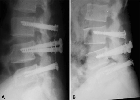
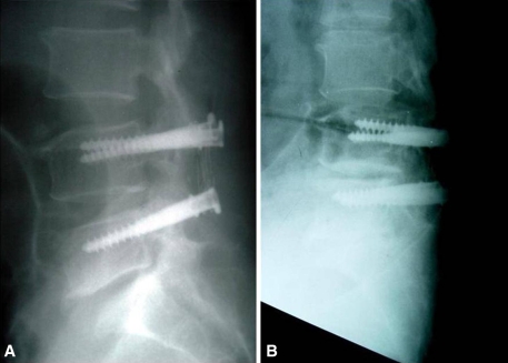
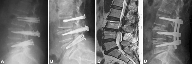
Although there has been some enthusiasm over the early clinical results obtained using the Graf ligament, associated mid- to long-term results are controversial. We retrospectively reviewed 43 patients (67 segments) treated with the Graf ligament for degenerative lumbar stenosis. The minimum followup was 8 years (mean, 10 years; range, 8-14 years). At last followup, we observed angular instability in 19 of the 67 segments (28%) and translational instability in five (7%). The disc height decreased from postoperatively (mean 93% of the preoperative disc) to the final followup (mean 82%). Of the 43 patients, 18 (42%) had adjacent segmental instability at the upper segment, including angular instability in 11 patients, translational instability in four patients, and both in three patients. The adjacent segment instability at the lower segment revealed 13 patients (30%) with angular instability. The data suggest the anticipated mechanical effects of the Graf ligament can be altered by degeneration of the disc and facet joints at instrumented segments and the adjacent segment can be affected, perhaps as a result of abnormal load transmission.
Level of evidence: Level IV, therapeutic study.
Figures
References
-
- {'text': '', 'ref_index': 1, 'ids': [{'type': 'PubMed', 'value': '10505503', 'is_inner': True, 'url': 'https://pubmed.ncbi.nlm.nih.gov/10505503/'}]}
- Agazzi S, Reverdin A, May D. Posterior lumbar interbody fusion with cages: an independent review of 71 cases. J Neurosurg. 1999;91:186–192. - PubMed
-
- {'text': '', 'ref_index': 1, 'ids': [{'type': 'DOI', 'value': '10.1097/00007632-200111150-00004', 'is_inner': False, 'url': 'https://doi.org/10.1097/00007632-200111150-00004'}, {'type': 'PubMed', 'value': '11707702', 'is_inner': True, 'url': 'https://pubmed.ncbi.nlm.nih.gov/11707702/'}]}
- Ariga K, Miyamoto S, Nakase T, Okuda S, Meng W, Yonenobu K, Yoshikawa H. The relationship between apoptosis of endplate chondrocytes and aging and degeneration of the intervertebral disc. Spine. 2001;26:2414–2420. - PubMed
-
- {'text': '', 'ref_index': 1, 'ids': [{'type': 'DOI', 'value': '10.1007/BF01676569', 'is_inner': False, 'url': 'https://doi.org/10.1007/bf01676569'}, {'type': 'PMC', 'value': 'PMC3454634', 'is_inner': False, 'url': 'https://pmc.ncbi.nlm.nih.gov/articles/PMC3454634/'}, {'type': 'PubMed', 'value': '9093822', 'is_inner': True, 'url': 'https://pubmed.ncbi.nlm.nih.gov/9093822/'}]}
- Boos N, Webb JK. Pedicle screw fixation in spinal disorders: a European view. Eur Spine J. 1997;6:2–18. - PMC - PubMed
-
- {'text': '', 'ref_index': 1, 'ids': [{'type': 'DOI', 'value': '10.1097/00002517-199306060-00001', 'is_inner': False, 'url': 'https://doi.org/10.1097/00002517-199306060-00001'}, {'type': 'PubMed', 'value': '8130395', 'is_inner': True, 'url': 'https://pubmed.ncbi.nlm.nih.gov/8130395/'}]}
- Bridwell KH, Sedgewick TA, O’Brien MF, Lenke LG, Baldus C. The role of fusion and instrumentation in the treatment of degenerative spondylolisthesis with spinal stenosis. J Spinal Disord. 1993;6:461–472. - PubMed
-
- {'text': '', 'ref_index': 1, 'ids': [{'type': 'DOI', 'value': '10.1097/BRS.0b013e31814b2d8e', 'is_inner': False, 'url': 'https://doi.org/10.1097/brs.0b013e31814b2d8e'}, {'type': 'PubMed', 'value': '17873819', 'is_inner': True, 'url': 'https://pubmed.ncbi.nlm.nih.gov/17873819/'}]}
- Cheh G, Bridwell KH, Lenke LG, Buchowski JM, Daubs MD, Kim Y, Baldus C. Adjacent segment disease following lumbar/thoracolumbar fusion with pedicle screw instrumentation: a minimum 5-year follow-up. Spine. 2007;32:2253–2257. - PubMed
MeSH terms
LinkOut - more resources
Full Text Sources
Medical
Research Materials
