. 2009 Oct;11(5):1051-7.
doi: 10.1007/s10544-009-9322-8. Epub 2009 May 19.

A physiologically realistic in vitro model of microvascular networks

Affiliations

A physiologically realistic in vitro model of microvascular networks

Jenna M Rosano et al. Biomed Microdevices. 2009 Oct.

Abstract

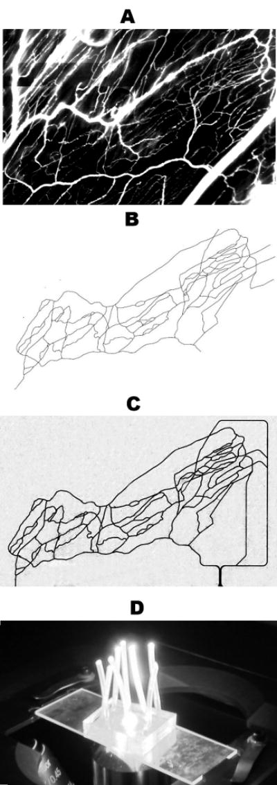
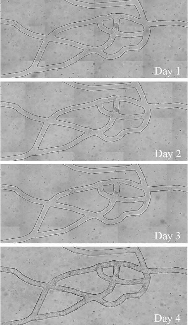
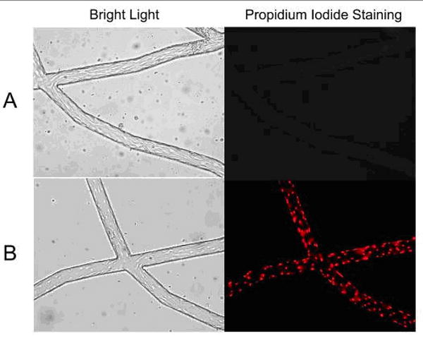
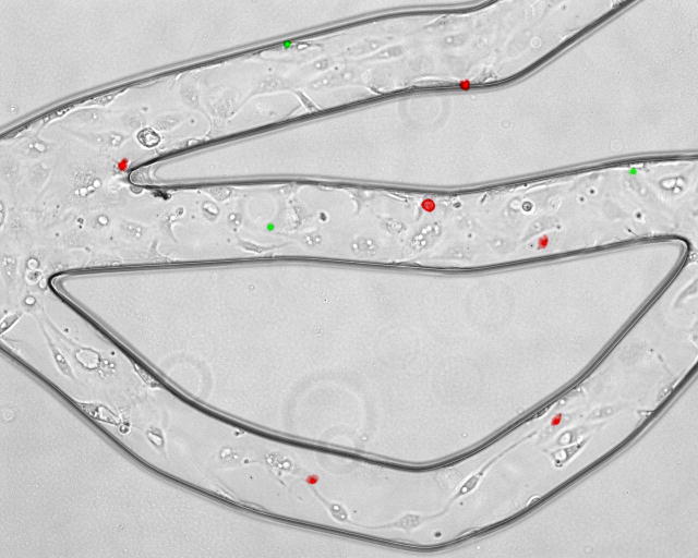
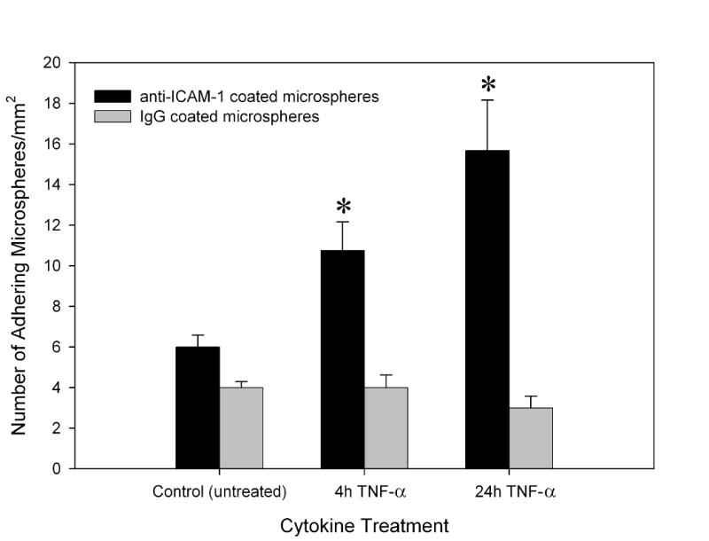
Existing microfluidic devices, e.g. parallel plate flow chambers, do not accurately depict the geometry of microvascular networks in vivo. We have developed a synthetic microvascular network (SMN) on a polydimethalsiloxane (PDMS) chip that can serve as an in vitro model of the bifurcations, tortuosities, and cross-sectional changes found in microvascular networks in vivo. Microvascular networks from a cremaster muscle were mapped using a modified Geographical Information System, and then used to manufacture the SMNs on a PDMS chip. The networks were cultured with bovine aortic endothelial cells (BAEC), which reached confluency 3-4 days after seeding. Propidium iodide staining indicated viable and healthy cells showing normal behavior in these networks. Anti-ICAM-1 conjugated 2-mum microspheres adhered to BAEC cells activated with TNF-alpha in significantly larger numbers compared to control IgG conjugated microspheres. This preferential adhesion suggests that cultured cells retain an intact cytokine response in the SMN. This microfluidic system can provide novel insight into characterization of drug delivery particles and dynamic flow conditions in microvascular networks.


Figures

Figure 1
Figure 1
Panel A shows a montage of fluorescent images obtained in vivo from the cremaster muscle after I.V. injection of FITC-dextran. Our GIS-based system was used to digitize this microvascular network, panel B. This digitized map was then used to generate the synthetic microvascular network on PDMS, shown here perfused with trypan blue, panel C. The final “microvascular network on a chip” is shown in panel D.
Figure 2
Figure 2
Bovine aortic endothelial cells seeded in the PDMS chambers became confluent throughout the network after 3 to 4 days. Cells exhibited the normal cobblestone configuration in the direction of flow.
Figure 3
Figure 3
Endothelial cells cultured in the synthetic microvascular network were stained with propidium iodide (PI) to examine cell viability. A: The left panel demonstrates representative phase image of endothelial cells cultured in the SMN. Live endothelial cells cultured in the SMN were stained with PI at confluency. The right panel illustrates exclusion of PI in these cells, demonstrating their viability. B: The left panel shows representative phase images of control endothelial cells following fixation with ethanol. The right panel represents uptake of PI in positive controls, following fixation/permeabolization of these cells with ethanol.
Figure 4
Figure 4
Anti-ICAM-1 (red) and IgG1 (green) conjugated microspheres attached to activated bovine aortic endothelial cells in one region of the network. Anti-ICAM-1 conjugated microspheres show preferential adhesion to endothelial cells as compared to IgG conjugated microspheres.
Figure 5
Figure 5
Anti-ICAM-1 conjugated microspheres were found to adhere preferentially to bovine aortic endothelial cells within the synthetic microvascular network (SMN) after 4 and 24 hour activation with TNF-α. Mean ± SEM; n=4 SMN per TNF-α treated groups and n=3 SMN for untreated control group, * P < 0.05 indicates significant differences in anti-ICAM-1 adhesion in SMN compared to the non-activated control group.

References

    1. Camp JP, Stokol T, Shuler ML. Biomed Microdevices. 2007;10(2) PMCID: 17891456. - PubMed
    1. Caputo KE, Lee D, King MR, Hammer DA. Biophys J. 2007;92(3) PMCID: 17085490. - PMC - PubMed
    1. Cokelet GR, Soave R, Pugh G, Rathbun L. Microvasc Res. 1993;46(3) PMCID: 8121322. - PubMed
    1. Decuzzi P, Ferrari M. Biomaterials. 2008;29(3) PMCID: 17936897. - PubMed
    1. El Ali J, Sorger PK, Jensen KF. Nature. 2006;442(7101) PMCID: 16871208. - PubMed

Publication types