

Aberrant trafficking of human melanocortin 1 receptor variants associated with red hair and skin cancer: Steady-state retention of mutant forms in the proximal golgi
- PMID: 19452503
- PMCID: PMC2705480
- DOI: 10.1002/jcp.21804
Aberrant trafficking of human melanocortin 1 receptor variants associated with red hair and skin cancer: Steady-state retention of mutant forms in the proximal golgi
Abstract
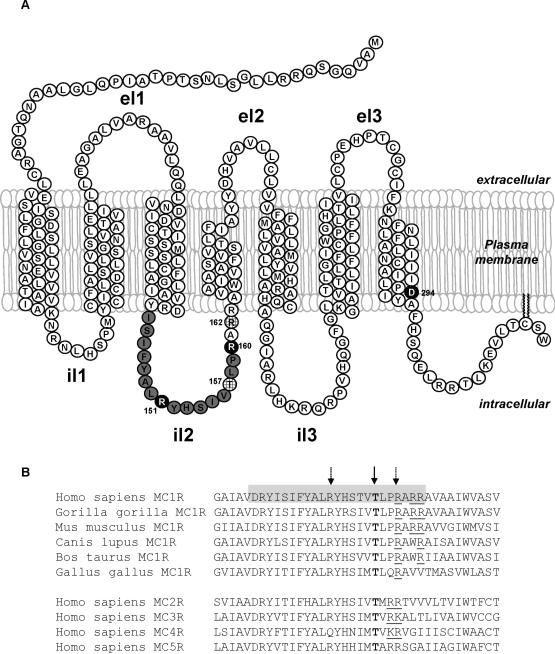
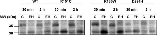
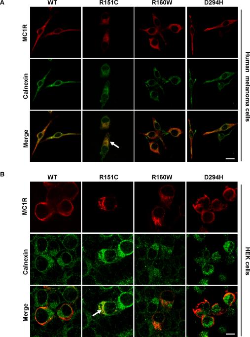
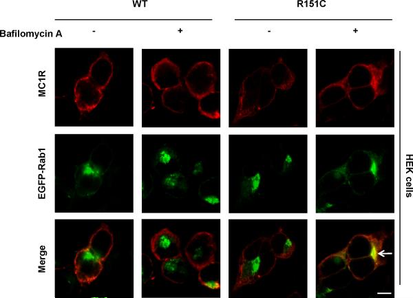
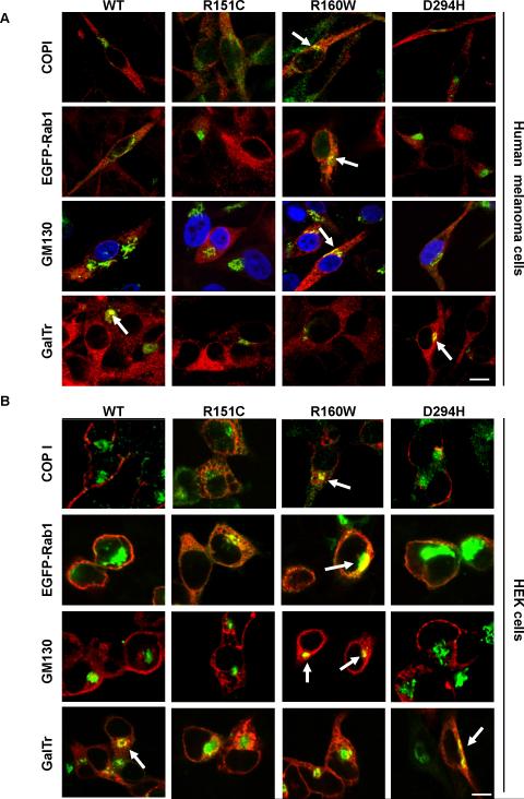
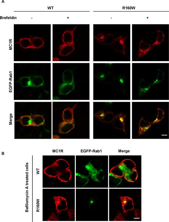
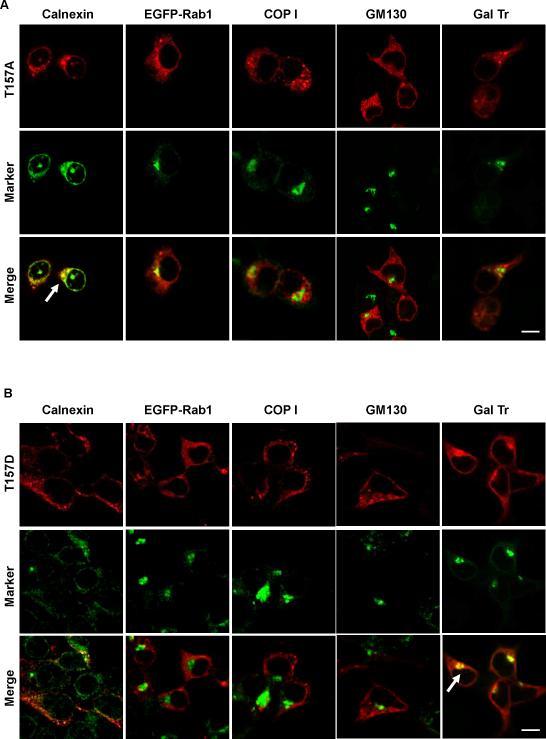
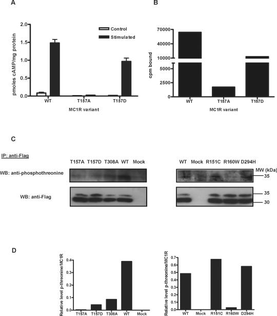
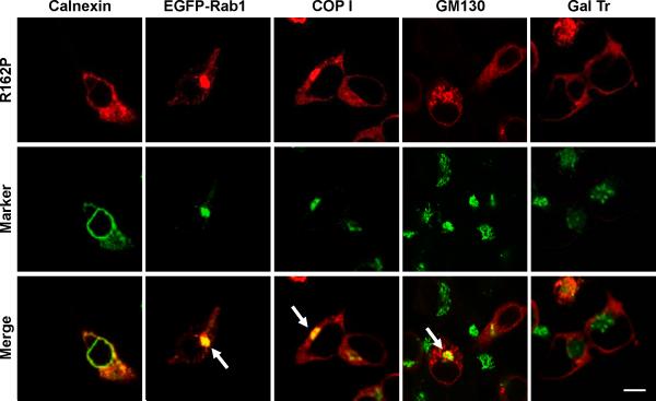
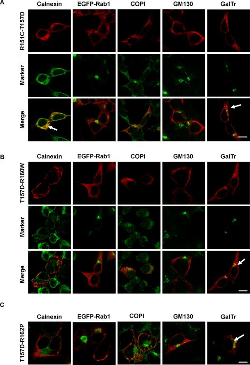
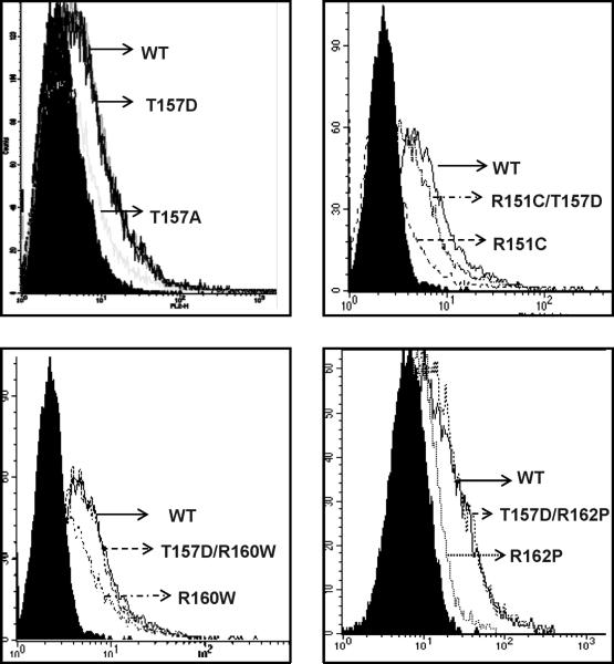
The melanocortin 1 receptor (MC1R), a Gs protein-coupled receptor (GPCR) expressed in melanocytes, is a major determinant of skin pigmentation and phototype. MC1R activation stimulates melanogenesis and increases the ratio of black, strongly photoprotective eumelanins to reddish, poorly photoprotective pheomelanins. Several MC1R alleles are associated with red hair, fair skin, increased sensitivity to ultraviolet radiation (the RHC phenotype) and increased skin cancer risk. Three highly penetrant RHC variants, R151C, R160W, and D294H are loss-of-function MC1R mutants with altered cell surface expression. In this study, we show that forward trafficking was normal for D294H. Conversely, export traffic was impaired for R151C, which accumulated in the endoplasmic reticulum (ER), and for R160W, which was enriched in the cis-Golgi. This is the first report of steady-state retention in a post-ER secretory compartment of a GPCR mutant found in the human population. Residues R151 and R160 are located in the MC1R second intracellular loop (il2). Two other mutations in il2, T157A preventing T157 phosphorylation and R162P disrupting a (160)RARR(163) motif, also caused intracellular retention. Moreover, T157 was phosphorylated in wild-type MC1R and a T157D mutation mimicking constitutive phosphorylation allowed normal traffic, and rescued the retention phenotype of R160W and R162P. Therefore, MC1R export is likely regulated by T157 phosphorylation and the (160)RARR(163) arginine-based motif functions as an ER retrieval signal. These elements are conserved in mammalian MC1Rs and in all five types of human melanocortin receptors. Thus, members of this GPCR subfamily might share common mechanisms for regulation of plasma membrane expression.
Figures
Similar articles
-
Regulation of human melanocortin 1 receptor signaling and trafficking by Thr-308 and Ser-316 and its alteration in variant alleles associated with red hair and skin cancer.J Biol Chem. 2007 Feb 2;282(5):3241-51. doi: 10.1074/jbc.M606865200. Epub 2006 Nov 27. J Biol Chem. 2007. PMID: 17130136
-
Altered cell surface expression of human MC1R variant receptor alleles associated with red hair and skin cancer risk.Hum Mol Genet. 2005 Aug 1;14(15):2145-54. doi: 10.1093/hmg/ddi219. Epub 2005 Jun 22. Hum Mol Genet. 2005. PMID: 15972726
-
Identification and functional analysis of novel variants of the human melanocortin 1 receptor found in melanoma patients.Hum Mutat. 2009 May;30(5):811-22. doi: 10.1002/humu.20971. Hum Mutat. 2009. PMID: 19338054
-
MC1R, the cAMP pathway, and the response to solar UV: extending the horizon beyond pigmentation.Pigment Cell Melanoma Res. 2014 Sep;27(5):699-720. doi: 10.1111/pcmr.12257. Epub 2014 May 30. Pigment Cell Melanoma Res. 2014. PMID: 24807163 Free PMC article. Review.
-
The melanocortin 1 receptor and the UV response of human melanocytes--a shift in paradigm.Photochem Photobiol. 2008 Mar-Apr;84(2):501-8. doi: 10.1111/j.1751-1097.2008.00294.x. Epub 2008 Feb 11. Photochem Photobiol. 2008. PMID: 18282187 Review.
Cited by
-
Reciprocal responses of fibroblasts and melanocytes to α-MSH depending on MC1R polymorphisms.Dermatoendocrinol. 2011 Oct;3(4):259-65. doi: 10.4161/derm.3.4.17454. Epub 2011 Oct 1. Dermatoendocrinol. 2011. PMID: 22259655 Free PMC article.
-
A single lys residue on the first intracellular loop modulates the endoplasmic reticulum export and cell-surface expression of α2A-adrenergic receptor.PLoS One. 2012;7(12):e50416. doi: 10.1371/journal.pone.0050416. Epub 2012 Dec 5. PLoS One. 2012. PMID: 23227171 Free PMC article.
-
A naturally occurring GIP receptor variant undergoes enhanced agonist-induced desensitization, which impairs GIP control of adipose insulin sensitivity.Mol Cell Biol. 2014 Oct 1;34(19):3618-29. doi: 10.1128/MCB.00256-14. Epub 2014 Jul 21. Mol Cell Biol. 2014. PMID: 25047836 Free PMC article.
-
How Genes Modulate Patterns of Aging-Related Changes on the Way to 100: Biodemographic Models and Methods in Genetic Analyses of Longitudinal Data.N Am Actuar J. 2016;20(3):201-232. doi: 10.1080/10920277.2016.1178588. Epub 2016 Jun 22. N Am Actuar J. 2016. PMID: 27773987 Free PMC article.
-
Identification and functional characterization of natural human melanocortin 1 receptor mutant alleles in Pakistani population.Pigment Cell Melanoma Res. 2015 Nov;28(6):730-5. doi: 10.1111/pcmr.12400. Epub 2015 Sep 22. Pigment Cell Melanoma Res. 2015. PMID: 26197705 Free PMC article.
References
-
- Ahner A, Brodsky JL. Checkpoints in ER-associated degradation: excuse me, which way to the proteasome? Trends Cell Biol. 2004;14(9):474–478. - PubMed
-
- Beaumont KA, Newton RA, Smit DJ, Leonard JH, Stow JL, Sturm RA. Altered cell surface expression of human MC1R variant receptor alleles associated with red hair and skin cancer risk. Hum Mol Genet. 2005;14(15):2145–2154. - PubMed
-
- Beaumont KA, Shekar SN, Newton RA, James MR, Stow JL, Duffy DL, Sturm RA. Receptor function, dominant negative activity and phenotype correlations for MC1R variant alleles. Hum Mol Genet. 2007;16(18):2249–2260. - PubMed
-
- Bohm M, Luger TA, Tobin DJ, Garcia-Borron JC. Melanocortin receptor ligands: new horizons for skin biology and clinical dermatology. J Invest Dermatol. 2006;126(9):1966–1975. - PubMed
Publication types
MeSH terms
Substances
Grants and funding
LinkOut - more resources
Full Text Sources
Medical
Molecular Biology Databases
