. 2009 Jul;238(7):1788-95.
doi: 10.1002/dvdy.21977.

Zebrafish wnt3 is expressed in developing neural tissue

Affiliations

Zebrafish wnt3 is expressed in developing neural tissue

Wilson K Clements et al. Dev Dyn. 2009 Jul.

Abstract

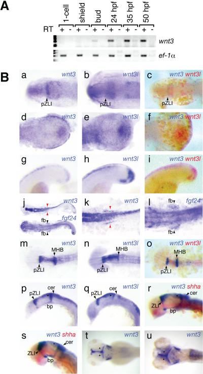
Wnt signaling regulates embryonic patterning and controls stem cell homeostasis, while aberrant Wnt activity is associated with disease. One Wnt family member, Wnt3, is required in mouse for specification of mesoderm, and later regulates neural patterning, apical ectodermal ridge formation, and hair growth. We have identified and performed preliminary characterization of the zebrafish wnt3 gene. wnt3 is expressed in the developing tailbud and neural tissue including the zona limitans intrathalamica (ZLI), optic tectum, midbrain-hindbrain boundary, and dorsal hindbrain and spinal cord. Expression in these regions suggests that Wnt3 participates in processes such as forebrain compartmentalization and regulation of tectal wiring topography by retinal ganglia axons. Surprisingly, wnt3 expression is not detectable during mesoderm specification, making it unlikely that Wnt3 regulates this process in zebrafish. This lack of early expression should make it possible to study later Wnt3-regulated patterning events, such as neural patterning, by knockdown studies in zebrafish.


Figures

Fig. 1
Fig. 1
Zebrafish wnt3 is the orthologue of other vertebrate Wnt3 genes. A: Sequence conservation of the deduced amino acid sequence for zebrafish Wnt3, comparing mouse Wnt3 (mWnt3, top), human WNT3 (hWNT3, second from top), zebrafish Wnt3l (zWnt3l, second from bottom), and mouse Wnt3a (mWnt3a) bottom. B: Phylogenetic analysis of zWnt3 compared to similar genes in zebrafish mouse and human. zWnt3 clusters with known human and mouse Wnt3 genes, whereas zwnt3l clusters with mWnt3a. mWnt8a was used as the outgroup. Numbers indicate bootstrap confidences at branch points. C: Conservation of synteny between Wnt3 genes in human, mouse, fugu, and zebrafish. The Nsf gene is the nearest 3'-neighbor in all four species.
Fig. 2
Fig. 2
Temporal and spatial expression of wnt3 during zebrafish embryonic development. A: RT-PCR analysis of wnt3 (top row) in total cDNA from whole embryos at the indicated stages of development. ef-1α was used as a positive control. B: WISH analysis of wnt3 expression. Embryos at various ages processed for expression of the mRNAs indicated. In all cases anterior is to the left. Flatmounted head views at 3-s. (a–c) show that wnt3 displays stronger expression in the presumptive ZLI (black arrows, pZLI), whereas wnt3l displays stronger expression in the prospective midbrain just posterior. Flatmounted tailbud views at 3-s. (d–f). Note wnt3 shows stronger expression in the lateral and posterior border of the tailbud. Lateral views of the tailbud at 18-s. (g–i). wnt3l shows stronger and deeper expression in the tailbud. Comparative flatmount views of wnt3 and fgf24 at 22-s. (j) and magnified images at the level of the finbud progenitors (k, l). wnt3 is expressed bilaterally (red arrowheads) in tissue just medial to the finbud progenitor fields marked by fgf24 (black arrowheads, fb). Dorsal flatmount views at 18-s. (m–o). Both wnt3 and wnt3l are expressed in the developing diencephalon and mesencephalon, but wnt3 is expressed farther posterior into the presumptive cerebellum. Lateral views of the brainstem with eyes removed at 18-s. (p–r) confirm wnt3 expression into the presumptive cerebellum and demonstrate that wnt3 extends much farther ventrally at both the presumptive ZLI and the midbrain hindbrain boundary, reaching into the presumptive basal plate. At 24 and 30 hpf (r, s; lateral brain stem views, eyes removed), this trend continues. Double WISH with shha confirms the basal plate expression. At 4 dpf (t) and 6 dpf (u) wnt3 continues to be expressed in the optic tectum, and is visible in the otic vesicles (dorsal head views). Anatomical landmarks: bp, basal plate; cer, cerebellum; fb, finbud progenitors; MHB, midbrain-hindbrain boundary; pZLI, presumptive zona limitans intrathalamica; ZLI, zona limitans intrathalamica; * optic tectum.
Fig. 3
Fig. 3
Zebrafish Wnt3 can activate the canonical Wnt pathway. Zebrafish heterozygous for a gfp transgene under the control of a Wnt/β-catenin-dependent promoter (Tg(TOP:GFP)w25/+) were outcrossed, and the resulting embryos were allowed to develop uninjected (left column), or injected with either 30 pg (middle column) or 70pg (right column) of wnt3 mRNA. At shield stage, embryos were fixed and processed by WISH for expression of either gfp (top row) or chd (bottom row) as indicated. Injected embryos showed a dose-dependent increase in canonical Wnt signaling-driven transcript abundance. 100% of embryos displayed the phenotypes depicted for each condition. Non-transgenic siblings provided an internal control for gfp in situs (not pictured).

References

    1. Barrow JR, Howell WD, Rule M, Hayashi S, Thomas KR, Capecchi MR, McMahon AP. Wnt3 signaling in the epiblast is required for proper orientation of the anteroposterior axis. Dev Biol. 2007;312:312–320. - PubMed
    1. Barrow JR, Thomas KR, Boussadia-Zahui O, Moore R, Kemler R, Capecchi MR, McMahon AP. Ectodermal Wnt3/beta-catenin signaling is required for the establishment and maintenance of the apical ectodermal ridge. Genes Dev. 2003;17:394–409. - PMC - PubMed
    1. Bovolenta P, Rodriguez J, Esteve P. Frizzled/RYK mediated signalling in axon guidance. Development. 2006;133:4399–4408. - PubMed
    1. Braun MM, Etheridge A, Bernard A, Robertson CP, Roelink H. Wnt signaling is required at distinct stages of development for the induction of the posterior forebrain. Development. 2003;130:5579–5587. - PubMed
    1. Buckles GR, Thorpe CJ, Ramel MC, Lekven AC. Combinatorial Wnt control of zebrafish midbrain-hindbrain boundary formation. Mech Dev. 2004;121:437–447. - PubMed

Publication types

MeSH terms

LinkOut - more resources