. 2009 Nov 1;77(2):359-69.
doi: 10.1002/prot.22441.

Engineered cystine knot peptides that bind alphavbeta3, alphavbeta5, and alpha5beta1 integrins with low-nanomolar affinity

Affiliations

Engineered cystine knot peptides that bind alphavbeta3, alphavbeta5, and alpha5beta1 integrins with low-nanomolar affinity

Richard H Kimura et al. Proteins. .

Abstract

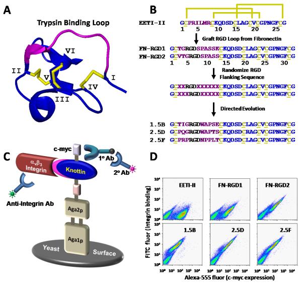
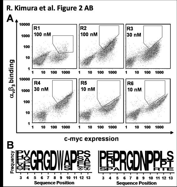
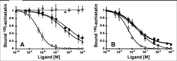
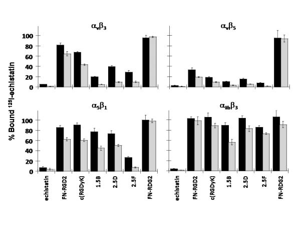
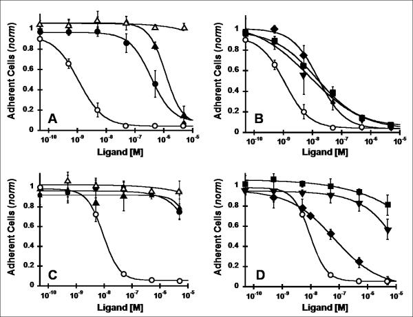
There is a critical need for compounds that target cell surface integrin receptors for applications in cancer therapy and diagnosis. We used directed evolution to engineer the Ecballium elaterium trypsin inhibitor (EETI-II), a knottin peptide from the squash family of protease inhibitors, as a new class of integrin-binding agents. We generated yeast-displayed libraries of EETI-II by substituting its 6-amino acid trypsin binding loop with 11-amino acid loops containing the Arg-Gly-Asp integrin binding motif and randomized flanking residues. These libraries were screened in a high-throughput manner by fluorescence-activated cell sorting to identify mutants that bound to alpha(v)beta(3) integrin. Select peptides were synthesized and were shown to compete for natural ligand binding to integrin receptors expressed on the surface of U87MG glioblastoma cells with half-maximal inhibitory concentration values of 10-30 nM. Receptor specificity assays demonstrated that engineered knottin peptides bind to both alpha(v)beta(3) and alpha(v)beta(5) integrins with high affinity. Interestingly, we also discovered a peptide that binds with high affinity to alpha(v)beta(3), alpha(v)beta(5), and alpha(5)beta(1) integrins. This finding has important clinical implications because all three of these receptors can be coexpressed on tumors. In addition, we showed that engineered knottin peptides inhibit tumor cell adhesion to the extracellular matrix protein vitronectin, and in some cases fibronectin, depending on their integrin binding specificity. Collectively, these data validate EETI-II as a scaffold for protein engineering, and highlight the development of unique integrin-binding peptides with potential for translational applications in cancer.


Figures

Fig. 1
Fig. 1
EETI-II engineering by yeast surface display. (A) Three dimensional structure of EETI-II (pdb: 2it7). The three disulfide bonds that stabilize the core of the molecule are shown in yellow. Individual cysteine residues are numbered I-VI. (B) Strategy for EETI-II engineering. The primary structure of EETI-II shows the characteristic disulfide bonds of cysteines 1 – 6 (yellow). The sequences of three representative mutants isolated from the first (1.5B) and second (2.5D, 2.5F) rounds of directed evolution are shown. (C) Schematic of the yeast display system. Expression of EETI-II mutants as Aga2p fusions is measured by flow cytometry through detection of a C-terminal c-myc epitope tag using a chicken anti-c-myc antibody and an Alexa 555-labeled secondary antibody. Integrin binding is measured with a FITC-labeled anti-integrin antibody. (D) Flow cytometry dot plots showing c-myc expression (x-axis) and αvβ3 integrin binding (y-axis) of peptides displayed on the surface of yeast.
Fig. 2
Fig. 2
Library sort progressions and sequences from the second round of directed evolution. (A) Density dot plots indicating FITC (y-axis) and Alexa-555 (x-axis) fluorescence on a single-cell level. Yeast-displayed knottin libraries were screened by dual-color FACS for mutants that bound the highest levels of αvβ3 integrin for a given amount of expression. Six rounds of FACS were used to obtain an enriched population of yeast that displayed clones with high αvβ3 integrin binding affinity. Integrin concentrations were reduced in successive rounds of sorting from 100 nM (rounds 1-2), to 30 nM (rounds 3-4), and 10 nM (rounds 5-6). (B) Sequence logos showing the relative frequencies of amino acids present in the engineered EETI-II loop from 22 individual clones. Two distinct families of binders emerged from library sorting and are represented by 17 clones (left) and 5 clones (right). The first amino acid shown is the third amino acid of the parent scaffold EETI-II. This figure was generated using online weblogo software (weblogo.berkeley.edu).
Fig. 3
Fig. 3
Competition binding of peptides to tumor cell surface integrins. Varying concentrations of unlabeled peptides were incubated with 125I-labeled echistatin and allowed to compete for binding to integrin receptors expressed on the surface of U87MG glioblastoma cells. The fraction of 125I-echistatin bound to the cell surface is plotted versus the concentration of unlabeled (A) c(RGDyK) (●), FN-RGD2 (▲), and FN-RDG2 (△), and (B) evolved mutants 1.5B (▼), 2.5D (■), and 2.5F (◆). (A,B) Unlabeled echistatin (○) was used as a positive control to compare binding data from different experiments. Data shown is the average of triplicate values and error bars represent standard deviations. IC50 values are shown in Table I.
Fig. 4
Fig. 4
Competition binding of peptides to surface-immobilized integrins. To determine integrin binding specificity, 125I-labeled echistatin and 5 nM (black bars) or 50 nM (grey bars) unlabeled peptides were added to microtiter plates coated with detergent-solubilized integrin receptor subtypes αvβ3, αvβ5, α5β1, and αiibβ3. Unbound 125I-echistatin was removed, and the amount of plate-bound 125I-echistatin remaining was measured. Unlabeled echistatin, which binds to all four integrins with high affinity, was used as a positive control. Error bars represent the standard deviation of measurements performed in triplicate.
Fig. 5
Fig. 5
Inhibition of integrin-dependent cell adhesion. Vitronectin (A,B) or fibronectin (C,D) coated strips were incubated with U87MG cells for 2 h with the indicated concentrations of peptides. Adherent cells remaining after several wash steps were quantified with crystal violet staining by absorbance at 600 nm. Values were normalized to a negative control containing no competing peptide. Symbols are: (A,C) c(RGDyK) (●), FN-RGD2 (▲), and FN-RDG2 (△), and (B,D) evolved mutants 1.5B (▼), 2.5D (■), and 2.5F (◆). Unlabeled echistatin (○) was used as a positive control to compare cell adhesion data from different experiments. Data shown is the average of triplicate values and error bars represent standard deviations. IC50 values are shown in Table I.

References

    1. Folkman J. The role of angiogenesis in tumor growth. Semin Cancer Biol. 1992;3:65–71. - PubMed
    1. Alghisi GC, Ruegg C. Vascular integrins in tumor angiogenesis: mediators and therapeutic targets. Endothelium. 2006;13:113–135. - PubMed
    1. Brooks PC, Clark RA, Cheresh DA. Requirement of vascular integrin alpha v beta 3 for angiogenesis. Science. 1994;264:569–571. - PubMed
    1. Kim S, Bell K, Mousa SA, Varner JA. Regulation of angiogenesis in vivo by ligation of integrin alpha5beta1 with the central cell-binding domain of fibronectin. Am J Pathol. 2000;156:1345–1362. - PMC - PubMed
    1. Mizejewski GJ. Role of integrins in cancer: survey of expression patterns. Proc Soc Exp Biol Med. 1999;222:124–138. - PubMed

Publication types

MeSH terms

LinkOut - more resources