Review
. 2009:10:153-74.
doi: 10.1146/annurev-genom-082908-150150.

The posttranslational processing of prelamin A and disease

Affiliations
Review

The posttranslational processing of prelamin A and disease

Brandon S J Davies et al. Annu Rev Genomics Hum Genet. 2009.

Abstract

Human geneticists have shown that some progeroid syndromes are caused by mutations that interfere with the conversion of farnesyl-prelamin A to mature lamin A. For example, Hutchinson-Gilford progeria syndrome is caused by LMNA mutations that lead to the accumulation of a farnesylated version of prelamin A. In this review, we discuss the posttranslational modifications of prelamin A and their relevance to the pathogenesis and treatment of progeroid syndromes.


Figures

Figure 1
Figure 1
Biogenesis of lamin A and the failure to generate mature lamin A in the setting of ZMPSTE24 deficiency (restrictive dermopathy) and HGPS. Prelamin A (664 amino acids) normally undergoes four posttranslational processing steps (left panel). First, the cysteine of the CaaX motif is farnesylated by FTase. Second, the –aaX is released. Third, the newly exposed farnesylcysteine is methylated. Fourth, the carboxyl-terminal 15 amino acids, including the farnesylcysteine methyl ester, are clipped off by ZMPSTE24 and degraded, releasing mature lamin A (646 amino acids). In the setting of ZMPSTE24 deficiency (middle panel), the last endoproteolytic processing step does not occur, resulting in the accumulation of the farnesylated form of prelamin A. In the setting of HGPS (right panel), a point mutation results in a 50–amino acid deletion in prelamin A (amino acids 607–656), which removes the site for the second endoproteolytic cleavage. Thus, the farnesylated mutant prelamin A (progerin) accumulates in cells, and no mature lamin A is formed. Modified, with permission, from the Journal of Lipid Research (117).
Figure 2
Figure 2
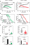
Phenotypic comparison of LmnanHG/+ mice and FTI-treated LmnaHG/+ mice. When compared to Lmna+/+ and LmnaHG/+ mice, FTI-treated LmnaHG/+ mice (left, green) and LmnanHG/+ mice (right, red) have very similar body weight curves (a), survival patterns (b), number of rib fractures (c), and body fat (d). Modified, with permission, from The Journal of Clinical Investigation (112, 114) and Biochimica et Biophysica Acta (116).

References

    1. Ackerman J, Gilbert-Barness E. Hutchinson-Gilford progeria syndrome: a pathologic study. Pediatr. Pathol. Mol. Med. 2002;21:1–13. - PubMed
    1. Agarwal AK, Fryns J-P, Auchus RJ, Garg A. Zinc metalloproteinase, ZMPSTE24, is mutated in mandibuloacral dysplasia. Hum. Mol. Genet. 2003;12:1995–2001. - PubMed
    1. Batstone MD, Macleod AWG. Oral and maxillofacial surgical considerations for a case of Hutchinson-Gilford progeria. Int. J. Paediatr. Dent. 2002;12:429–32. - PubMed
    1. Beck LA, Hosick TJ, Sinensky M. Incorporation of a product of mevalonic acid metabolism into proteins of Chinese hamster ovary cell nuclei. J. Cell Biol. 1988;107:1307–16. - PMC - PubMed
    1. Beck LA, Hosick TJ, Sinensky M. Isoprenylation is required for the processing of the lamin A precursor. J. Cell Biol. 1990;110:1489–99. - PMC - PubMed

Publication types

LinkOut - more resources