. 2009 Jul;39(7):618-25.
doi: 10.1111/j.1365-2362.2009.02153.x. Epub 2009 May 6.

SARS-coronavirus modulation of myocardial ACE2 expression and inflammation in patients with SARS

Affiliations

SARS-coronavirus modulation of myocardial ACE2 expression and inflammation in patients with SARS

G Y Oudit et al. Eur J Clin Invest. 2009 Jul.

Abstract

Background: Angiotensin converting enzyme 2 (ACE2), a monocarboxylase that degrades angiotensin II to angiotensin 1-7, is also the functional receptor for severe acute respiratory syndrome (SARS) coronavirus (SARS-CoV) and is highly expressed in the lungs and heart. Patients with SARS also suffered from cardiac disease including arrhythmias, sudden cardiac death, and systolic and diastolic dysfunction.

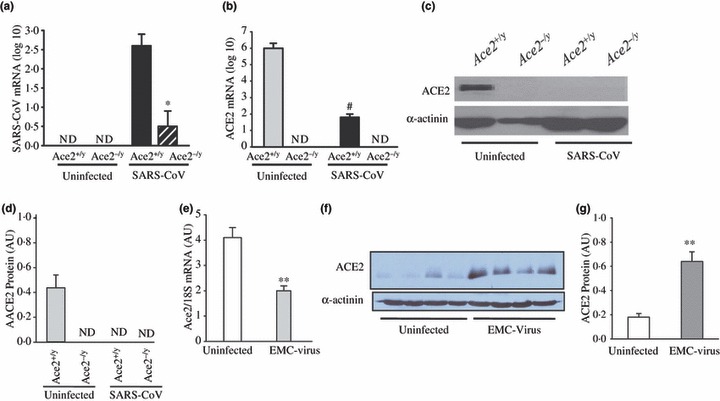
Materials and methods: We studied mice infected with the human strain of the SARS-CoV and encephalomyocarditis virus and examined ACE2 mRNA and protein expression. Autopsy heart samples from patients who succumbed to the SARS crisis in Toronto (Canada) were used to investigate the impact of SARS on myocardial structure, inflammation and ACE2 protein expression.

Results: Pulmonary infection with the human SARS-CoV in mice led to an ACE2-dependent myocardial infection with a marked decrease in ACE2 expression confirming a critical role of ACE2 in mediating SARS-CoV infection in the heart. The SARS-CoV viral RNA was detected in 35% (7/20) of autopsied human heart samples obtained from patients who succumbed to the SARS crisis during the Toronto SARS outbreak. Macrophage-specific staining showed a marked increase in macrophage infiltration with evidence of myocardial damage in patients who had SARS-CoV in their hearts. The presence of SARS-CoV in the heart was also associated with marked reductions in ACE2 protein expression.

Conclusions: Our data show that SARS-CoV can mediate myocardial inflammation and damage associated with down-regulation of myocardial ACE2 system, which may be responsible for the myocardial dysfunction and adverse cardiac outcomes in patients with SARS.


Figures

Figure 1
Figure 1
Pulmonary SARS‐CoV infection leads to myocardial SARS‐CoV infection and down‐regulation of myocardial ACE2 expression. (a–d) Human SARS‐CoV mRNA in the hearts of infected mice showing a clear dependence on ACE2 for myocardial SARS‐CoV infection (a) with down‐regulation of myocardial Ace2 mRNA expression based on real‐time PCR (b) and myocardial ACE2 protein levels shown by Western blot analysis (c) and quantification (d) in response to pulmonary SARS‐CoV infection. *P <0·01 compared with infected Ace2+/y group; #P <0·01 compared with uninfected group, n = 5, ND, not detectable. (e–g) Discordant changes in myocardial Ace2 mRNA and myocardial ACE2 protein levels in encephalomyocarditis (EMC) virus‐induced myocarditis with real‐time PCR showing reduced myocardial Ace2 mRNA (e) with increased myocardial ACE2 protein levels based on Western blot analysis (f) and quantification (g). **P <0·01 compared with placebo group, n = 5.
Figure 2
Figure 2
Detection of SARS‐CoV genome in postmortem human heart samples with evidence of myocardial inflammation and damage. (a) Presence of SARS‐CoV genome in the heart of 35% of the patients who died from SARS (+SARS‐CoV, open bar, n = 7) and its negative impact on illness duration compared with patients who died from SARS without SARS‐CoV in the heart (‐SARS‐CoV, closed bar, n = 13); #P <0·05 compared with +SARS‐CoV group. (b–h) Representative trichrome‐stained myocardial section obtained from a patient who died from non‐SARS related sepsis (bacterial pneumonia) (b), SARS with evidence of SARS‐CoV in the heart (c), SARS without evidence of SARS‐CoV in the heart (d) showing increased interstitial fibrosis and inflammation (e) and cardiomyocyte hypertrophy based on myocyte cross‐sectional area (MCSA) (f) without evidence of apoptosis in patients who died from SARS with (g) and without (h) evidence of SARS‐CoV in the heart. Scale bar represents 50 μM. *P <0·01 compared with all other groups. VL‐ve and VH‐ve = patients who died from a non‐SARS related sepsis (open bar), VL+ve and VH+ve = patients who died from SARS with SARS‐CoV in the heart (grey bar) and VL+ve and VH‐ve = patients who died from SARS without SARS‐CoV in the heart (closed bar), n = 7 per group.
Figure 3
Figure 3
Increased macrophage infiltration in the absence of increased lymphocytic infiltration in the left ventricle of patients who died from SARS. (a–e) Representative anti‐CD68‐stained immunohistochemistry section from a patient who died from non‐SARS related sepsis (bacterial pneumonia) (a), SARS with evidence of SARS‐CoV in the heart (b), SARS without evidence of SARS‐CoV in the heart (c) and a positive control section obtained from human spleen (d) with quantification of myocardial macrophage count (e). (f–i) Representative anti‐CD3 immunohistochemistry illustrating a representative section from a patient who died from SARS with evidence of SARS‐CoV in the heart (f), SARS without evidence of SARS‐CoV in the heart (g) and a positive control section obtained from human spleen (h) with quantification of myocardial lymphocyte count (i). Scale bar represents 50 μM. #P <0·05 compared with all other groups. VL‐ve and VH‐ve = patients who died from a non‐SARS related sepsis (open bar), VL+ve and VH+ve = patients who died from SARS with SARS‐CoV in the heart (grey bar) and VL+ve and VH‐ve = patients who died from SARS without SARS‐CoV in the heart (closed bar), n = 7 per group.
Figure 4
Figure 4
Reduced ACE2 protein expression in patients who died from SARS and had SARS‐CoV detected in their hearts. (a–e) Representative sections showing staining for ACE2 in the heart from a patient who died from non‐SARS related sepsis (bacterial pneumonia) (a), SARS with evidence of SARS‐CoV in the heart (b), SARS without evidence of SARS‐CoV in the heart (c), a negative control section (d) while pre‐incubation with recombinant human ACE2 (1 mg mL−1) prevented ACE2 staining (e). Scale bar represents 50 μM. (f) Quantification of ACE2 immunohistochemical staining showing reduced ACE2 protein expression in patients who died from SARS with SARS‐CoV in their hearts. *P <0·05 compared with all other groups; VL‐ve and VHve = patients who died from a non‐SARS related sepsis (open bar), VL+ve and VH+ve = patients who died from SARS with SARS‐CoV in the heart (grey bar) and VL+ve and VH‐ve = patients who died from SARS without SARS‐CoV in the heart (closed bar), n = 7 per group.

Similar articles

Cited by

References

    1. Crackower MA, Sarao R, Oudit GY, Yagil C, Kozieradzki I, Scanga SE et al. Angiotensinconverting enzyme 2 is an essential regulator of heart function. Nature 2002;417:822–8. - PubMed
    1. Oudit GY, Crackower MA, Backx PH, Penninger JM. The role of ACE2 in cardiovascular physiology. Trends Cardiovasc Med 2003;13:93–101. - PubMed
    1. Oudit GY, Kassiri Z, Patel MP, Chappell M, Butany J, Backx PH et al. Angiotensin II‐mediated oxidative stress and inflammation mediate the age‐dependent cardiomyopathy in ACE2 null mice. Cardiovasc Res 2007;75:29–39. - PubMed
    1. Oudit GY, Herzenberg AM, Kassiri Z, Wong D, Reich H, Khokha R et al. Loss of angiotensinconverting enzyme‐2 leads to the late development of angiotensin II‐dependent glomerulosclerosis. Am J Pathol 2006;168:1808–20. - PMC - PubMed
    1. Imai Y, Kuba K, Rao S, Huan Y, Guo F, Guan B et al. Angiotensin‐converting enzyme 2 protects from severe acute lung failure. Nature 2005;436:112–6. - PMC - PubMed

Publication types