

Mucinous cystic neoplasms of the mesentery: a case report and review of the literature
- PMID: 19454018
- PMCID: PMC2691402
- DOI: 10.1186/1477-7819-7-47
Mucinous cystic neoplasms of the mesentery: a case report and review of the literature
Abstract
Background: Mucinous cystic neoplasms arise in the ovary and various extra-ovarian sites. While their pathogenesis remains conjectural, their similarities suggest a common pathway of development. There have been rare reports involving the mesentery as a primary tumour site.
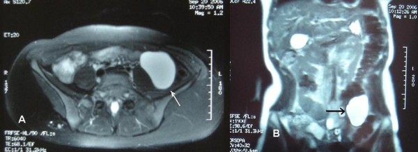
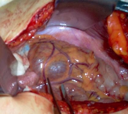
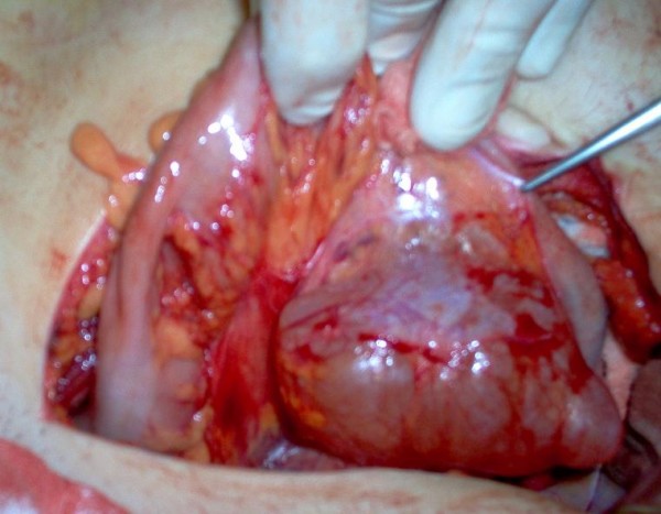
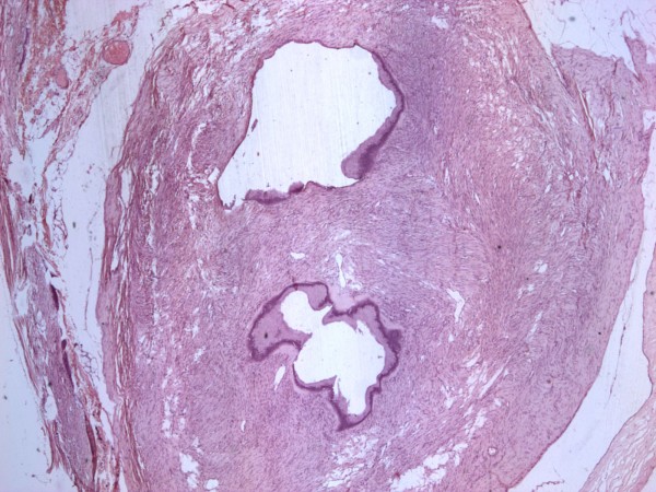
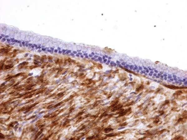
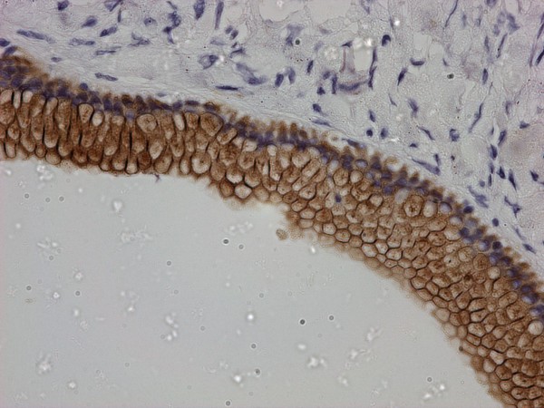
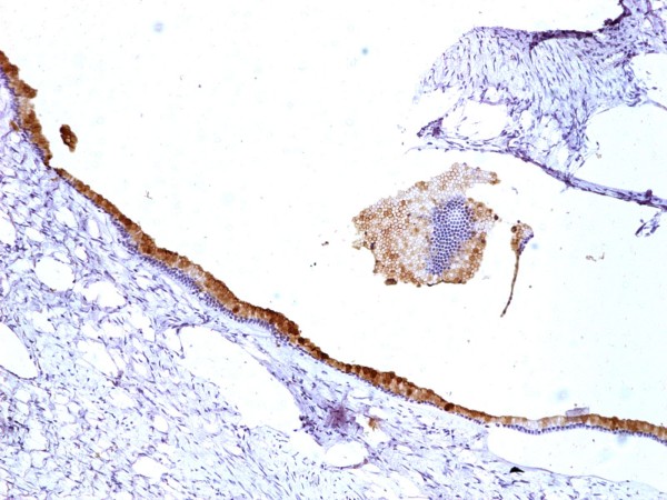
Case presentation: A cystic mass of uncertain origin was demonstrated radiologically in a 22 year old female with chronic abdominal pain. At laparotomy, the mass was fixed within the colonic mesentery. Histology demonstrated a benign mucinous cystadenoma.
Methods and results: We review the literature on mucinous cystic neoplasms of the mesentery and report on the pathogenesis, biologic behavior, diagnosis and treatment of similar extra-ovarian tumors. We propose an updated classification of mesenteric cysts and cystic tumors.
Conclusion: Mucinous cystic neoplasms of the mesentery present almost exclusively in women and must be considered in the differential diagnosis of mesenteric tumors. Only full histological examination of a mucinous cystic neoplasm can exclude a borderline or malignant component. An updated classification of mesenteric cysts and cystic tumors is proposed.
Figures
References
-
- Vanek VW, Phillips AK. Retroperitoneal, mesenteric and omental cysts. Arch Surg. 1984;119:838–42. - PubMed
-
- Pisano G, Erdas E, Parodo G, et al. Acute abdomen due to rupture of mesenteric cysts. Observations on a clinical case and review of the literature. Minerva Chir . 2004;59:405–411. - PubMed
-
- Ekçi B, Ayan F, Gürses B. Ruptured mesenteric cyst: a rare presentation after trauma. Ulus Travma Acil Cerrahi Derg. 2007;13:74–7. - PubMed
Publication types
MeSH terms
LinkOut - more resources
Full Text Sources
