Review
. 2009 Apr 1;387(1):1-12.
doi: 10.1016/j.ab.2009.01.011. Epub 2009 Jan 19.

Direct detection and quantification of microRNAs

Affiliations
Review

Direct detection and quantification of microRNAs

Eric A Hunt et al. Anal Biochem. .

Abstract

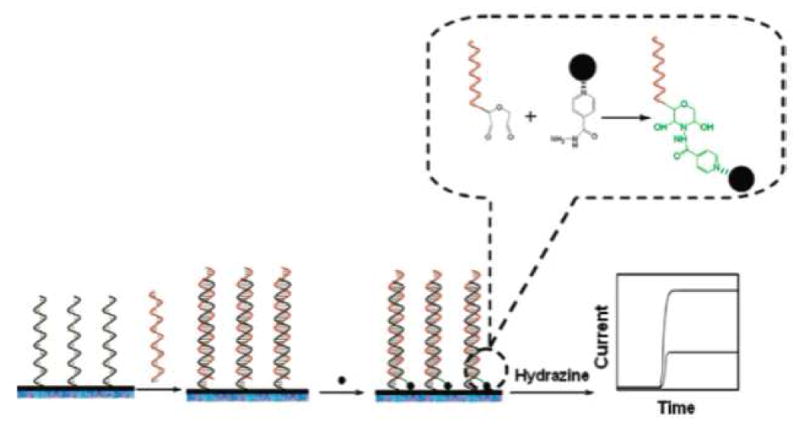
The recent discovery of the potent regulatory nature of microRNAs (miRNAs), a relatively new class of approximately 22 nucleotide RNAs, has made them a primary focus in today’s biochemical and medical research. The relationship between miRNA expression patterns and the onset of cancer, as well as other diseases, has glimpsed the potential of miRNAs as disease biomarkers or drug targets, making them a primary research focus. Their promising future in medicine is hinged upon improving our scientific understanding of their intricate regulatory mechanisms. In the realm of analytical chemistry, the main challenge associated with miRNA is its detection. Their extremely small size and low cellular concentration poses many challenges for achieving reliable results. Current reviews in this area have focused on adaptations to microarray, PCR, and Northern blotting procedures to make them suitable for miRNA detection. While these are extremely powerful methods and accepted as the current standards, they are typically very laborious, semi-quantitative, and often require expensive imaging equipment and/or radioactive/toxic labels. This review aims to highlight emerging techniques in miRNA detection and quantification that exhibit superior flexibility and adaptability as well as matched or increased sensitivity in comparison to the current standards. Specifically, this review will cover colorimetric, fluorescence, bioluminescence, enzyme, and electrochemical based methods, which drastically reduce procedural complexity and overall expense of operation thereby increasing the accessibility of this field of research. The methods are presented and discussed as to their improvements over current standard methods as well as their potential complications preventing acceptance as standard procedures. These new methods have addressed the many of the problems associated with miRNA detection through the employment of enzyme-based signal amplification, enhanced hybridization conditions using PNA capture probes, highly sensitive and flexible forms of spectroscopy, and extremely responsive electrocatalytic nanosystems, among other approaches.


Figures

Figure 1
Figure 1
The genes coding for the primary miRNA transcript are typically found in intergenic regions or within the introns of eukaryotic protein-coding genes. The primary transcript is further processed and capped for nuclear export to the cytoplasm where the precursor form is cleaved into the 18–24 nucleotide mature sequence, which is incorporated into the RISC protein complex. Figure adapted from Macmillan Publishers Ltd: H. Grosshans, and W. Filipowicz, Molecular biology: the expanding world of small RNAs. Nature 451 (2008) 414–6.
Figure 2
Figure 2
In the colorimetric assay designed by Yang and colleagues, the functionalized gold nanoparticle binds adjacent to the biotinylated probe on the miRNA target, thus allowing it to be immobilized on a streptavidin coated plate. The grayscale absorbance from the silver enhancement is used for colorimetric quantification of target. Figure adapted from Elsevier B.V.: W.J. Yang, X.B. Li, et al., Quantification of microRNA by gold nanoparticle probes. Analytical Biochemistry 376 (2008) 183–8.
Figure 3
Figure 3
In the FCS based assay developed by Neely and colleagues, free probes were sequestered by specially designed quenching probes. The red 2 laser was used to correlate the red 1 and green signals. Reprinted by permission from Macmillan Publishers Ltd: L. Neely, S. Patel, et al., A single-molecule method for the quantitation of microRNA gene expression. Nat Meth 3 (2006) 41–6.
Figure 4
Figure 4
In the molecular beacon assay developed by Paiboonskuwong, adjacent guanine in the primary and precursor forms of the target successfully quenches the fluorophore and limits signal production to the mature form only. Reprinted by permission from Oxford University Press: K. Paiboonskuwong, and Y. Kato, Detection of the mature, but not precursor, RNA using a fluorescent DNA probe. Nucleic Acids Symposium Series (2006) 327–8.
Figure 5
Figure 5
(A) The miRNA Invader assay developed by Allawi and colleagues is based on the same principles as the RNA Invader assay. (B) A general FRET oligo may be used for all sequences as it anneals to the SRT. (C) Self-complementary adaptations must be made to the invasive and probe oligos to compensate for the small size of the miRNA target. Reprinted by permission from the RNA Society: H.T. Allawi, J.E. Dahlberg, et al., Quantitation of microRNAs using a modified Invader assay. RNA 10 (2004) 1153–61.
Figure 6
Figure 6
In the absence of the target, utilized in the BRET-based assay developed by Cissell et al., the Rluc-probe and the QD-probe hybridize creating a BRET signal. In the presence of the target, there is a competition between the target and the Rluc-probe, decreasing the BRET signal. With kind permission from Springer Science+Business Media: Bioanal Chem, Rapid single-step nucleic acid detection, 319, 2008, 2577, K.A. Cissell, Y. Rahimi, S. Shrestha, E.A. Hunt, and S.K. Deo, fig. 1.
Figure 7
Figure 7
The miRNA (both labeled and unlabeled) hybridize to the biotinylated probe, in the solid phase assay described by Cissell et al. The hybridized probe is immobilized onto a neutravidin plate, washed to remove unbound miRNA, and the emission signal recorded. Figure adapted from the American Chemical Society: K.A. Cissell, Y. Rahimi, et al., Bioluminescence-based detection of microRNA mir21 in breast cancer cells. Anal Chem 80 (2008) 2319–25.
Figure 8
Figure 8
In the enzymatic/colorimetric assay developed by Su and colleagues, the PNA capture probe improves hybridization conditions and localizes negative charge for HRP adsorption. Reprinted by permission from the American Chemical Society: X. Su, H.F. Teh, et al., Enzyme-based colorimetric detection of nucleic acids using peptide nucleic acid-immobilized microwell plates. Anal Chem 79 (2007) 7192–7.
Figure 9
Figure 9
The catalytic activity of a ribozyme is directly related to its structural conformation. Reprinted by permission from Elsevier B.V.: M.J. Fedor, Structure and function of the hairpin ribozyme. J Mol Biol 297 (2000) 269–91.
Figure 10
Figure 10
In the ribozyme based assay developed by Hartig and colleagues, the stem-loop domain (C) prevents cleavage of the FRET oligo when not bound to target by restricting the ribozyme to an inactive conformation and blocking the substrate binding domain (A). Figure adapted from the American Chemical Society: J.S. Hartig, I. Grüne, et al., Sequence-specific detection of microRNAs by signal-amplifying ribozymes. J Am Chem Soc 126 (2004) 722–3.
Figure 11
Figure 11
In the electrochemical method developed by Gao et al, the miRNA was ligated, via a covalent bond, to (Os(dmpy)2(IN)Cl+) through a simple condensation reaction. Figure adapted from Elsevier B.V.: Z. Gao, and Y.H. Yu, A microRNA biosensor based on direct chemical ligation and electrochemically amplified detection. Sensors and Actuators B: Chemical 121 (2007) 552–9.
Figure 12
Figure 12
For the electrochemical work by Gao et al., the labeled miRNA was hybridized to the immobilized probes; the oxidation signal was recorded upon the addition of hydrazine. Figure adapted from Elsevier B.V.: Z. Gao, and Y.H. Yu, Direct labeling microRNA with an electrocatalytic moiety and its application in ultrasensitive microRNA assays. Biosensors and Bioelectronics 22 (2007) 933–40.
Figure 13
Figure 13
Nanoparticles were chemically ligated to the hybridized miRNAs, in the electrocatalytic assays developed by Gao et al., through a condensation reaction. Reprinted by permission from the American Chemical Society: Z. Gao, and Z. Yang, Detection of microRNAs using electrocatalytic nanoparticle tags. Anal Chem 78 (2006) 1470–7.
Figure 14
Figure 14
For the electrochemical assay developed by Fan and colleagues, the total RNA sample was hybridized to the capture probes, creating a net negative charge. The hybridized miRNA was then incubated with an aniline mixture. The protonated aniline molecules align around the hybridized miRNA forming polyaniline polymers. The nanowires were then expanded by doping with HCl vapors, creating an electrically conducting network bridging the gaps of the biosensor array. Reprinted by permission from the American Chemical Society: Y. Fan, X. Chen, et al., Detection of microRNAs using target-guided formation of conducting polymer nanowires in nanogaps. J Am Chem Soc 129 (2007) 5437–43.

References

    1. Lee RC, Feinbaum RL, Ambros V. The C. elegans heterochronic gene lin-4 encodes small RNAs with antisense complementarity to lin-14. Cell. 1993;75:843–54. - PubMed
    1. Reinhart BJ, Slack FJ, Basson M, Pasquinelli AE, Bettinger JC, Rougvie AE, Horvitz HR, Ruvkun G. The 21-nucleotide let-7 RNA regulates developmental timing in Caenorhabditis elegans. Nature. 2000;403:901–6. - PubMed
    1. Bartel DP. MicroRNAs: genomics, biogenesis, mechanism, and function. Cell. 2004;116:281–97. - PubMed
    1. Kim VN, Nam JW. Genomics of microRNA. Trends Genet. 2006 - PubMed
    1. Gregory RI, Shiekhattar R. MicroRNA biogenesis and cancer. Cancer Res. 2005;65:3509–12. - PubMed

Publication types

LinkOut - more resources