. 2009 May 8;5(5):476-86.
doi: 10.1016/j.chom.2009.03.011.

Lipocalin-2 resistance confers an advantage to Salmonella enterica serotype Typhimurium for growth and survival in the inflamed intestine

Affiliations

Lipocalin-2 resistance confers an advantage to Salmonella enterica serotype Typhimurium for growth and survival in the inflamed intestine

Manuela Raffatellu et al. Cell Host Microbe. .

Abstract

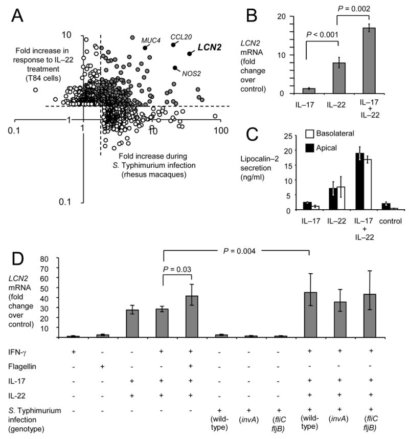
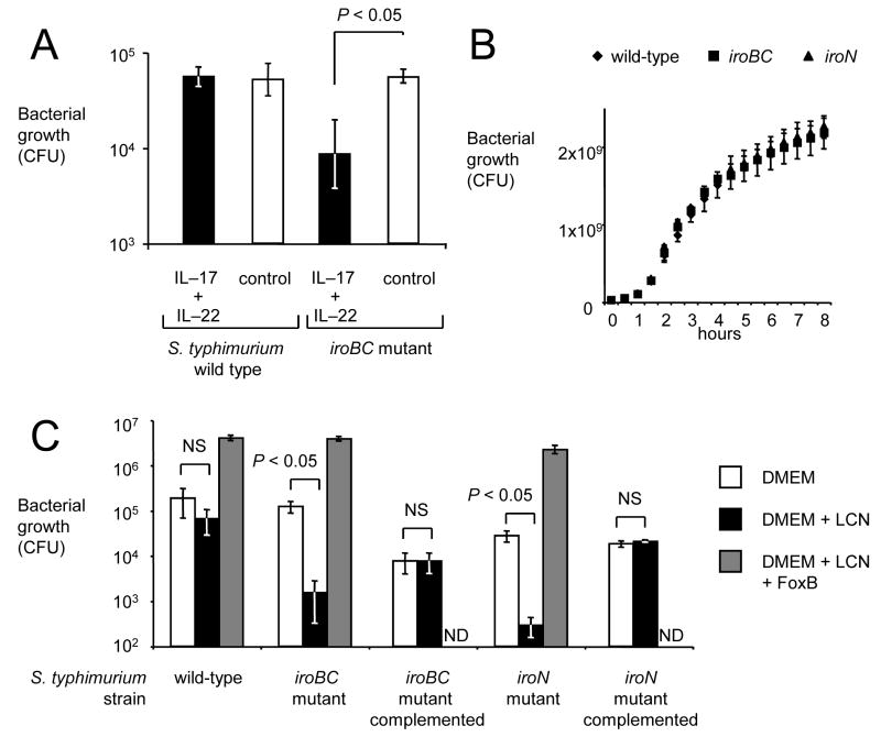
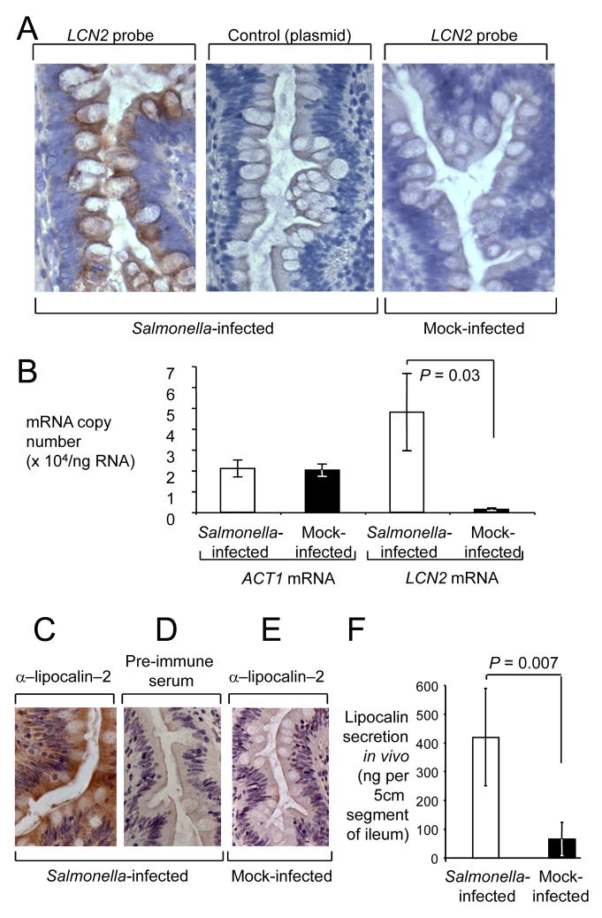
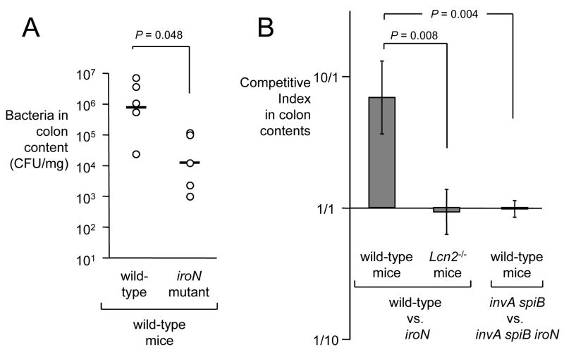
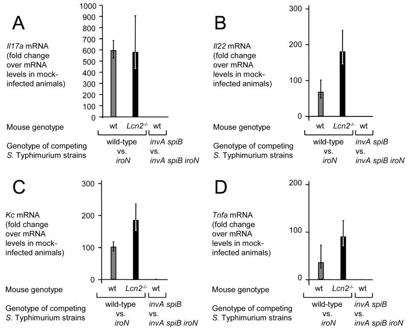
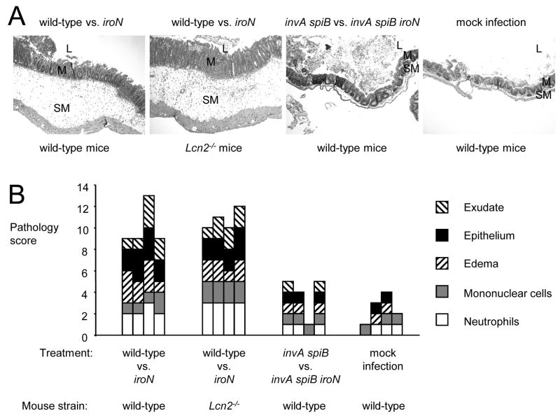
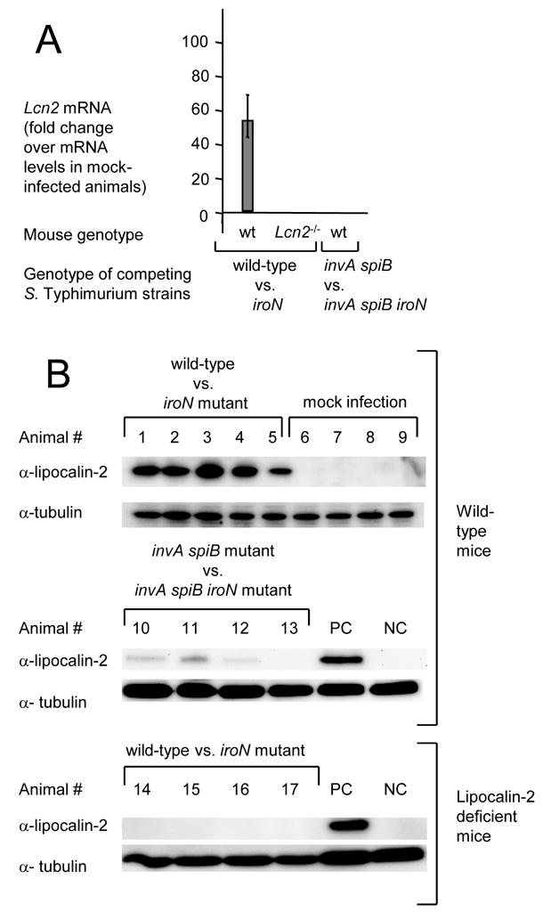
In response to enteric pathogens, the inflamed intestine produces antimicrobial proteins, a process mediated by the cytokines IL-17 and IL-22. Salmonella enterica serotype Typhimurium thrives in the inflamed intestinal environment, suggesting that the pathogen is resistant to antimicrobials it encounters in the intestinal lumen. However, the identity of these antimicrobials and corresponding bacterial resistance mechanisms remain unknown. Here, we report that enteric infection of rhesus macaques and mice with S. Typhimurium resulted in marked Il-17- and IL-22-dependent intestinal epithelial induction and luminal accumulation of lipocalin-2, an antimicrobial protein that prevents bacterial iron acquisition. Resistance to lipocalin-2, mediated by the iroBCDE iroN locus, conferred a competitive advantage to the bacterium in colonizing the inflamed intestine of wild-type but not of lipocalin-2-deficient mice. Thus, resistance to lipocalin-2 defines a specific adaptation of S. Typhimurium for growth in the inflamed intestine.


Figures

Figure 1
Figure 1
Expression of lipocalin–2 by polarized intestinal model epithelia upon stimulation with IL–17 and/or IL–22. (A) Meta analysis of the overlap (gray circles) between increases (>2 fold) in transcripts in the ileal mucosa of rhesus macaques during S. Typhimurium infection (x axis) and induction of gene expression observed in human T84 epithelial cells after treatment with IL–22 (y axis). The identity of selected genes (closed circles) is indicated. (B and D) Detection of LCN2 expression in polarized T84 cells stimulated for 4 hours with reagents listed below each graph using quantitative real-time PCR. (C) Secretion of lipocalin–2 by T84 cells into the apical (open bars) or basolateral (closed bars) compartment detected by ELISA. (B–D) Data represent means ± standard deviation from at least three different experiments.
Figure 2
Figure 2
The iroBC locus confers lipocalin-2 resistance by overcoming iron limitation. (A) Growth of the S. Typhimurium wild type or an isogenic iroBC mutant in medium from collected from polarized T84 cells that had either not been stimulated with cytokines (control, open bars) or had been stimulated with IL–17 and IL–22 for 24 hours (IL–17 + IL–22, closed bars). Bacterial numbers were determined 5 hours after inoculation with 103 bacteria. (B) Bacterial growth in rich medium (LB broth). (C) Bacterial growth in tissue culture medium (DMEM) supplemented with lipocalin-2 (LCN), ferrioxamine B (FoxB) or containing no supplements. ND, not determined. All data represent means ± standard deviation from at least three different experiments.
Figure 3
Figure 3
Spatial and quantitative analysis of lipocalin–2 expression in the ileal mucosa of rhesus macaques during S. Typhimurium infection. (A) Detection of LCN2 transcripts (brown precipitate) in representative sections from a S. Typhimurium–infected loop (left panel) and a mock–infected loop (right panel) from the same animal by in situ hybridization. Hybridization with the empty plasmid vector used for cloning the LCN2 probe was performed as a negative control (middle panel). All sections were counterstained with hematoxylin. (B) Absolute transcript levels of LCN2 and ACT1 in mock–infected (closed bars) and S. Typhimurium–infected loops (open bars) from 4 rhesus macaques. Data represent mean mRNA copy numbers per ng of RNA ± standard error. Statistically significant (P < 0.05) differences are indicated by P values. (C–E) Detection of lipocalin-2 (brown precipitate) in representative sections from a S. Typhimurium–infected loop (C and D) and a mock–infected loop (E) from the same animal using immunohistochemistry with rabbit anti–rhesus lipocalin–2 antiserum (α–lipocalin–2). Immunohistochemistry with pre–immune serum was performed as a negative control (D). (F) Lipocalin–2 secretion in mock–infected (closed bars) and S. Typhimurium-infected loops (open bars) from 4 rhesus macaques. Data represents the total amount of lipocalin–2 (ng) secreted into an approximately 5 cm long segment of the ileum. Statistically significant (P < 0.05) differences are indicated by P values.
Figure 4
Figure 4
Growth of iroN deficient and iroN proficient S. Typhimurium strains in the murine intestine in vivo. (A) Recovery of S. Typhimurium from colon contents 48 hours after infection of mice with the S. Typhimurium wild-type or an iroN mutant. Each circle represent bacterial numbers recovered from an individual animal. Bars indicate the geometric mean. (B) Recovery of S. Typhimurium 48 hours after competitive infection with the indicated bacterial strains. Bars indicate the average competitive index (i.e. ratio of wild-type/iroN or ratio of invA spiB/invA spiB iroN) of bacteria recovered from colon contents of wild-type mice or Lcn2−/− mice 48 hours after infection with a 1:1 mixture the respective S. Typhimurium strains. Data represent geometric means ± standard error.
Figure 5
Figure 5
Inflammatory responses elicited in the cecum of streptomycin-pretreated mice 48 hours after S. Typhimurium infection assessed by measuring cytokine transcription. Transcript levels of Il17a (A), Il22 (B), Kc (C) and Tnfa (D) in wild type mice (wt, gray bars) or lipocalin-2 deficient mice (Lcn2−/−, black bars) 48 hours after competitive infection (wild-type vs. iroN or invA spiB vs. invA spiB iroN) was measured by quantitative real-time PCR. Bars represent fold-changes (geometric means) in mRNA levels compared to a group of mock-infected wild type mice ± standard error.
Figure 6
Figure 6
Histopathology of the cecum from mock-infected mice, or mice infected with a mixture of the indicated S. Typhimurium strains. (A) Histopathological appearance of the cecum from representative animals in each group. All images were taken from hematoxylin and eosin stained cecal sections at the same magnification (100x). Note marked edema in the submucosa (SM) of mice infected with S. Typhimurium wild-type vs. iron mutant. M, mucosa; L, lumen. (B) Pathology score determined by blinded examination of cecal sections under high power magnification. Each bar represents an individual animal.
Figure 7
Figure 7
Lipocalin-2 expression detected by quantitative real-time PCR (A) or by Western blot (B) in the cecal mucosa of streptomycin-pretreated mice 48 hours after S. Typhimurium infection. (A) Bars represent fold-changes (geometric means) in mRNA levels compared a group of mock-infected wild type mice ± standard error. (B) Lipocalin-2 expression in protein extracts from the cecal mucosa of individual mice was detected with anti-mouse lipocalin-2 antibody (α-lipocalin-2). Detection of tubulin with anti-mouse tubulin antibody (α-tubulin) served as a loading control for each sample. PC, positive control sample from animal #3; NC, negative control sample from animal #8.

Comment in

  • A precious metal heist.
    Skaar EP. Skaar EP. Cell Host Microbe. 2009 May 8;5(5):422-4. doi: 10.1016/j.chom.2009.05.005. Cell Host Microbe. 2009. PMID: 19454345

References

    1. Ackermann M, Stecher B, Freed NE, Songhet P, Hardt WD, Doebeli M. Self-destructive cooperation mediated by phenotypic noise. Nature. 2008;454:987–990. - PubMed
    1. Aujla SJ, Chan YR, Zheng M, Fei M, Askew DJ, Pociask DA, Reinhart TA, McAllister F, Edeal J, Gaus K, et al. IL-22 mediates mucosal host defense against Gram-negative bacterial pneumonia. Nat Med. 2008;14:275–281. - PMC - PubMed
    1. Ausubel FM, Brent R, Kingston RE, Moore DD, Seidman JG, Smith JA, Struhl K. Current protocols in molecular biology. J. Wiley & Sons; 1994.
    1. Barman M, Unold D, Shifley K, Amir E, Hung K, Bos N, Salzman N. Enteric salmonellosis disrupts the microbial ecology of the murine gastrointestinal tract. Infect Immun. 2008;76:907–915. - PMC - PubMed
    1. Barthel M, Hapfelmeier S, Quintanilla-Martinez L, Kremer M, Rohde M, Hogardt M, Pfeffer K, Russmann H, Hardt WD. Pretreatment of mice with streptomycin provides a Salmonella enterica serovar Typhimurium colitis model that allows analysis of both pathogen and host. Infect Immun. 2003;71:2839–2858. - PMC - PubMed

Publication types

MeSH terms