. 2009 Jun 1;182(11):6659-69.
doi: 10.4049/jimmunol.0804211.

Functionally distinct LAG-3 and PD-1 subsets on activated and chronically stimulated CD8 T cells

Affiliations

Functionally distinct LAG-3 and PD-1 subsets on activated and chronically stimulated CD8 T cells

Joseph F Grosso et al. J Immunol. .

Abstract

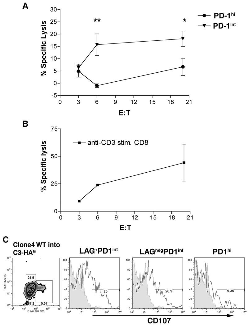
Lymphocyte Activation Gene-3 (LAG-3) is a transmembrane protein that binds MHC class II, enhances regulatory T cell activity, and negatively regulates cellular proliferation, activation, and homeostasis of T cells. Programmed Death 1 (PD-1) also negatively regulates T cell function. LAG-3 and PD-1 are both transiently expressed on CD8 T cells that have been stimulated during acute activation. However, both LAG-3 and PD-1 remain on CD8 T cells at high levels after stimulation within tolerizing environments. Our previous data demonstrated that blockade of either LAG-3 or PD-1 using mAb therapy in combination with vaccination restores the function of tolerized Ag-specific CD8 T cells in models of self and tumor tolerance. It is unclear whether tolerized CD8 T cells coexpress PD-1 and LAG-3 or whether PD-1 and LAG-3 mark functionally distinct populations of CD8 T cells. In this study, we describe three populations of CD8 T cells activated under tolerizing conditions based on LAG-3 and PD-1 staining, each with distinct phenotypic and functional characteristics. From a mechanistic perspective, both Ag concentration and proinflammatory signals control the expression of LAG-3 and PD-1 phenotypes on CD8 T cells under activating and tolerizing conditions. These results imply that signaling through the PD-1 and LAG-3 pathways have distinct functional consequences to CD8 T cells under tolerizing conditions and manipulation of both Ag and cytokine signaling can influence CD8 tolerance through LAG-3 and PD-1.


Figures

FIGURE 1
FIGURE 1
LAG-3 and PD-1 subsets have distinct phenotype and cytokine secretion patterns. A, Axial LN were harvested 3 days after transfer of Clone 4 CD8 T cells and single cell suspensions were stimulated in vitro with HA peptide and brefeldin A for 4 h. Cells were gated on Thy1.1+ cells to differentiate endogenous from transferred cells. Also shown are LAG-3 knockout Clone 4 CD8 cells as a gating control for LAG-3 as well as WT Clone 4 CD8 stained with LAG-3 and a PD-1 isotype Ab. B, IFN-γ and TNF-α analysis of LAG-3+PD-1int, LAG-3negPD-1int, and PD-1high CD8+Thy1.1+ populations after transfer into C3-HAhigh mice followed by in vitro stimulation. IFN-γ (C), TNF-α (D), ICOS (E), and 4-1BB (F) expression by CD8 subpopulations from C3-HAhigh axial lymph nodes. Shown are representative plots from one of four experiments each, involving three mice per group.
FIGURE 2
FIGURE 2
CD8 subsets differ in cytolytic ability. A, Thy1.1+Clone 4 CD8 T cells were transferred into Thy1.2+C3-HAhigh mice. After 3 days cells were sorted into CD8+Thy1.1+PD-1int or CD8+Thy1.1+PD-1high subsets and cultured in vitro for 3 days as described. B, Naive Clone 4 CD8 cells activated in vitro with anti-CD3 served as positive control killing. In vitro CTL function was assessed using HA-peptide pulsed target cells labeled with 51Cr. (**, p = 0.002; *, p = 0.04). Results shown are from one of three experiments. C, Expression of CD107 was determined on Clone 4 CD8 T cells transferred into C3-HAhigh mice after 3 days as described in Fig. 1.
FIGURE 3
FIGURE 3
CD8 subset differentiation occurs immediately after Ag exposure in vivo. A, Purified Clone 4 CD8+ cells (2 × 10⁶) were transferred into C3-HAhigh mice, and at the indicated times mice were sacrificed and axial LN were harvested for analysis. Single cell suspensions were stained for Thy1.1, LAG-3, and PD-1. B, After 4 days in C3-HAhigh mice, Clone 4 CD8 T cells were harvested and stimulated in vitro with HA peptide followed by Thy1.1, PD-1, LAG-3 and IFN-γ Ab staining. C, Ratio of LAG-3+PD-1int : LAG-3negPD-1int and LAG-3+PD-1high : LAG-3negPD-1high CD8 T cells as a function of time. D, Purified CFSE-labeled Thy1.1+ Clone 4 CD8 T cells were transferred into C3-HAhigh or C3-HAlow mice. After 2 days, axial LN were harvested and single cell suspensions were stained for Thy1.1, PD-1, LAG-3, and CFSE expression. Shown are the Thy1.1+ CFSE histograms. E, LAG-3 and PD-1 expression on Thy1.1+ T cells after adoptive transfer as a function of cell division. Each figure is representative of at least two independent experiments with similar results.
FIGURE 4
FIGURE 4
Ag concentration determines expression level of PD-1 and LAG-3 on HA-specific CD8 T cells. Thy1.2+C3-HAhigh and B10.d2 mice were adoptively transferred with 106 WT or LAG-3 knockout Thy1.1+ Clone 4 CD8 T cells and immediately vaccinated with an emulsion of IFA and varying concentrations of HA peptide (0, 5, 10, and 50 µg). Seven days after vaccination, vaccine-draining or contra-lateral lymph nodes were harvested, homogenized, and stained for Thy1.1, LAG-3, PD-1, and IFN-γ.
FIGURE 5
FIGURE 5
CD4 lymphocytes do not alter the expression of LAG-3 and PD-1 on CD8 cells. Thy1.1+Clone 4 CD8 cells (106) were transferred into C3-HAhigh (A) or GK1.5-treated C3-HAhigh (B) mice. Three days after transfer, Clone 4 CD8 cells were assessed for the expression of LAG-3, PD-1, and IFN-γ by flow cytometry.
FIGURE 6
FIGURE 6
TLR signaling reduces CD8 T cell expression of LAG-3 and enhances cell number and function. A, Thy1.2+C3-HAhigh mice were injected with LPS, poly(IC) i.p., or left untreated after adoptive transfer with 106 Thy1.1+ Clone 4 CD8. Mice received daily injections of either LPS or poly(IC). Five days after transfer, axial lymph nodes were harvested and assessed for Clone 4 CD8 expression of LAG-3 and CD62L. Top panels, Percent of Clone 4 CD8 cells in lymph nodes while bottom panels show the expression of LAG-3 and CD62L on Thy1.1+ cells. B, WT or LAG-3−/− Clone 4 CD8 cells were transferred into untreated, LPS, or poly(IC) injected C3-HAhigh mice. Five days after transfer, Thy1.1+CD8+ lymphocytes were sorted from spleen and lymph nodes and total RNA was prepared. The sorted populations were >98% pure. Real-time quantitative PCR for LAG-3 (C), ADAM10 (D), and ADAM17 (E). All samples are normalized to WT Clone 4 CD8 transferred into C3-HAhigh without TLR agonist treatment.

References

    1. Triebel F, Jitsukawa S, Baixeras E, Roman-Roman S, Genevee C, Viegas-Pequignot E, Hercend T. LAG-3, a novel lymphocyte activation gene closely related to CD4. J. Exp. Med. 1990;171:1393–1405. - PMC - PubMed
    1. Kisielow M, Kisielow J, Capoferri-Sollami G, Karjalainen K. Expression of lymphocyte activation gene 3 (LAG-3) on B cells is induced by T cells. Eur. J. Immunol. 2005;35:2081–2088. - PubMed
    1. Huang CT, Workman CJ, Flies D, Pan X, Marson AL, Zhou G, Hipkiss EL, Ravi S, Kowalski J, Levitsky HI, et al. Role of LAG-3 in regulatory T cells. Immunity. 2004;21:503–513. - PubMed
    1. Workman CJ, Cauley LS, Kim IJ, Blackman MA, Woodland DL, Vignali DA. Lymphocyte activation gene-3 (CD223) regulates the size of the expanding T cell population following antigen activation in vivo. J. Immunol. 2004;172:5450–5455. - PubMed
    1. Workman CJ, Vignali DA. The CD4-related molecule, LAG-3 (CD223), regulates the expansion of activated T cells. Eur. J. Immunol. 2003;33:970–979. - PubMed

Publication types

MeSH terms