. 2009 Jun 1;182(11):6697-708.
doi: 10.4049/jimmunol.0800997.

Chronic antigen stimulation alone is sufficient to drive CD8+ T cell exhaustion

Affiliations

Chronic antigen stimulation alone is sufficient to drive CD8+ T cell exhaustion

Christine M Bucks et al. J Immunol. .

Abstract

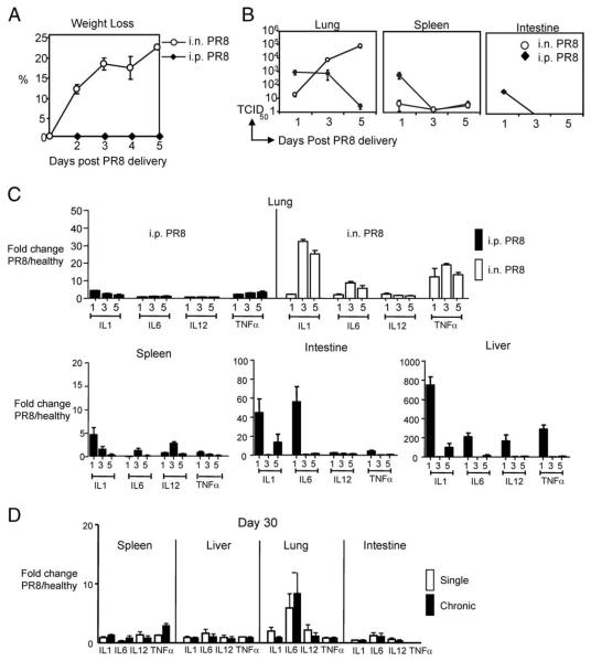
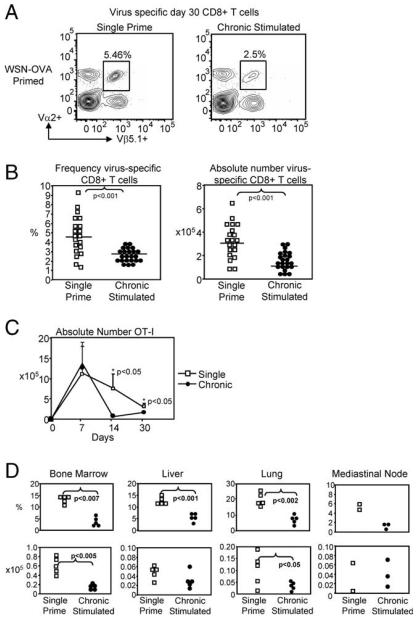
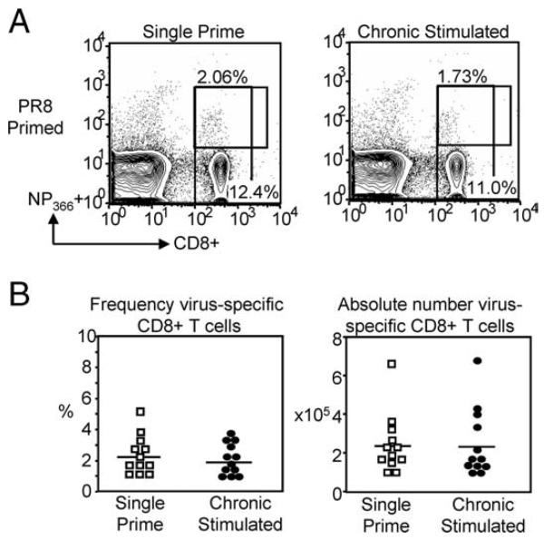
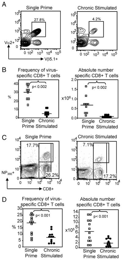
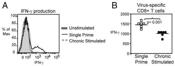
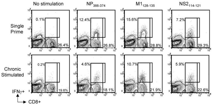
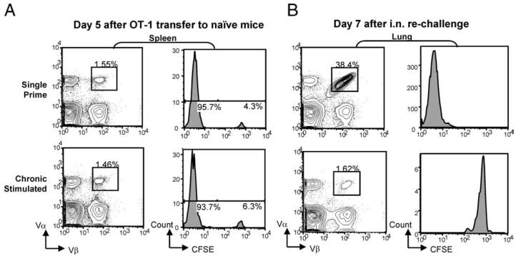
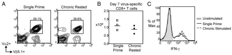
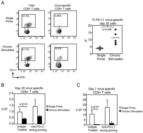
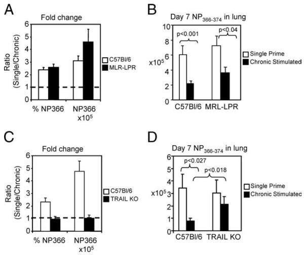
The failure of CD8(+) T cells to respond to chronic infection has been termed "exhaustion" and describes the condition in which CD8(+) T cells exhibit reduced differentiation, proliferation, and effector function. CD8(+) T cell exhaustion has been extensively studied in the murine model of chronic infection, lymphocytic choriomeningitis virus (LCMV). Although LCMV-based studies have yielded many interesting findings, they have not allowed for discrimination between the roles of cytokine- and Ag-driven exhaustion. We have created a system of chronic Ag stimulation using murine influenza A virus that leads to exhaustion and functional disability of virus-specific CD8(+) T cells, in the absence of high viral titers, sustained proinflammatory cytokine production and lymphocyte infection. Our findings show that Ag alone is sufficient to drive CD8(+) T cell impairment, that Ag-driven loss of virus-specific CD8(+) T cells is TRAIL mediated, and that removal of Ag reverses exhaustion. Although programmed death 1 was up-regulated on chronic Ag-stimulated CD8(+) T cells, it played no role in the exhaustion. These findings provide a novel insight into the mechanisms that control functional exhaustion of CD8(+) T cells in chronic infection.


Figures

FIGURE 1
FIGURE 1
The i.p. priming with PR8 did not lead to morbidity, systemic infection, cytokine burst, or chronic inflammation. C57BL/6 mice were i.n. infected or i.p. primed with 3 TCID50 or 3 × 10⁶ TCID50 influenza virus strain PR8. A, Weights of i.n. infected and i.p. primed mice were recorded daily (n = 5 mice/group). B, Viral load was measured by real-time PCR in the lung, spleen, kidney, and intestine on days 1, 3, and 5 after virus delivery (n = 3 mice/group for each time point). C, The level of mRNA was quantified, by real-time PCR, for IL-1, IL-6, IL-12, and TNF-α in the lung, spleen, intestine, and liver of i.p. primed or i.n. infected mice. D, Cytokine production (mRNA) quantified at day 30 after single or chronic Ag stimulation. Data are fold increase over healthy uninfected control lung mRNA (n = 3 mice/group per time point).
FIGURE 2
FIGURE 2
Chronic Ag stimulation reduced the quantity of day-30 virus-specific CD8+ T cells. OT-I transferred C57BL/6 mice were single or chronic stimulated with WSN-OVA. On day 30, the frequency of virus-specific CD8+ T cells was determined by flow cytometry. A, Representative flow cytometry virus-specific CD8+ T cells (gated on CD8+ T cells). B, Reduction in virus-specific CD8+ T cells was observed in pooled frequency and absolute number (n = 15 mice/group, p < 0.001). C, Kinetic analysis of virus-specific OT-I cells during 30 days of priming (n = 3 mice/group). D, Reduction of chronic CD8+ T cells in lymphoid and nonlymphoid tissues indicates migration is not impaired.
FIGURE 3
FIGURE 3
Chronic Ag stimulation of endogenous CD8+ T cells with PR8 did not significantly reduce the day-30 population. C57BL/6 mice received either a single i.p. injection or chronic Ag stimulation with influenza A virus strain PR8. After 30 days of stimulation, spleens were harvested and the frequency and absolute number of NP366-positive CD8+ T cells was assessed by multicolor flow cytometry. A, Representative FACS plots (left) show that there was no significant reduction in the frequency of day-30 virus-specific CD8+ T cells in chronic Ag-stimulated mice compared with single prime controls. Pooled frequency (right) indicates a trend toward a decrease in chronic stimulated CD8+ T cells, though no significance is reached. B, Pooled absolute numbers indicate that independent of chronic Ag stimulation the number of day 30 virus-specific CD8+ T cells remains comparable.
FIGURE 4
FIGURE 4
Chronic Ag stimulation reduced the quality of virus-specific CD8+ T cells. An equal number of single or chronic Ag-stimulated day-30 virus-specific CD8+ T cells were transferred to naive C57BL/6 mice. Recipient mice were i.n. challenged with 40 TCID50 WSN-OVA and harvested at the peak of the secondary response. Representative flow cytometry plots (A) and pooled frequencies and absolute numbers (B), showing a significant decrease in virus-specific CD8+ T cells in the lung of chronic mice compared with single prime mice. Each symbol represents a single animal. Representative flow cytometry plots (C) and pooled frequencies and absolute numbers (D) show that chronic Ag stimulation with PR8 also results in a significant reduction in responding virus-specific CD8+ T cells relative to single prime controls, following i.n. viral rechallenge.
FIGURE 5
FIGURE 5
Chronic Ag-stimulated day 7 secondary effector CD8+ T cells produced less IFN-γ. IFN-γ production by chronic Ag-stimulated virus-specific CD8+ T cells was defective compared with controls, after ex vivo stimulation with OVA257–264 peptide. A, Representative histogram (gated on CD8+ T cells) shows the reduction in IFN-γ production by chronic Ag-stimulated CD8+ T cells relative to single prime controls. B, Each symbol represents a single animal, pooled MFI of IFN-γ-producing cells (gated on CD8+ T cells) indicates decreased function of chronic Ag-stimulated CD8+ T cells compared with single prime controls (n = 6 mice, p < 0.001).
FIGURE 6
FIGURE 6
Chronic stimulation resulted in a loss of dominant and subdominant epitope responses. C57BL/6 mice received either a single i.p. prime or chronic stimulation for 30 days with 3 × 10⁶ TCID50 PR8. An equal number of single or chronic stimulated virus-specific CD8+ T cells were transferred to naive mice. Recipient mice were i.n. rechallenged with x31 and harvested at the peak of the secondary response on day 7 (n = 3 mice/group). Pulmonary lymphocytes were stimulated ex vivo with peptides to dominant (NP366–374) or subdominant epitopes (M1128–135 or NS2114–121) for 6 h and intracellular stained for IFN-γ production. Representative flow cytometry plots show that both dominant and subdominant epitope specific responses were reduced in chronic mice (bottom) compared with single prime controls (top).
FIGURE 7
FIGURE 7
Comparable survival of single and chronic stimulated CD8+ T cells after transfer to naive recipients. Day 30 single or chronic Ag-stimulated CD8+ T cells were CFSE-labeled and transferred to naive recipients. Recipients remained uninfected or were rechallenged i.n. with WSN-OVA virus. Mice were harvested on days 5 (healthy controls) and day 7 (virus-infected). A, Comparable frequency of CFSE-positive OT-I+ T cells were found in the spleens of naive recipients receiving either single or chronic stimulated CD8+ T cells. B, In the lungs of day 7 rechallenged mice, single prime CD8+ T cells exhibited robust expansion, resulting in an absence of CFSE-positive cells. Chronic Ag-stimulated CD8+ T cells failed to expand in number, and did not divide. Histograms depict CFSE stain on cells are gated on donor OT-I+ T cells based on Thy1.2 and Vα/Vβ expression.
FIGURE 8
FIGURE 8
Chronic Ag-stimulated virus-specific CD8+ T cells recovered function after Ag rest. A total of 1 × 10⁵ OT-I TCR Tg T cells were transferred to naive C57BL/6 mice. Mice were single or chronic stimulated with 1 × 10⁶ TCID50 WSN-OVA, by i.p. injection, for 30 days. On day 30, chronic animals were Ag rested for 45 days, and an equal number of single or chronic Ag-rested virus-specific CD8+ T cells were transferred to naive C57BL/6 mice, which were then i.n. challenged with WSN-OVA and harvested on day 7. A, Representative flow cytometry (gated on CD8+ T cells) from lungs of single or chronic rested infected mice is shown. As indicated by Vα2+Vβ5.1+ cells, chronic Ag-rested CD8+ T cells expanded equivalently to single prime animals. B, Pooled absolute numbers indicate that Ag rest recovers not only frequency, but also the number of chronic rested virus-specific CD8+ T cells (n = 3 mice/group). C, Representative flow cytometry histogram (gated on CD8+ T cells) shows equivalent MFI of IFN-γ production after ex vivo stimulation with OVA254–267-specific peptide.
FIGURE 9
FIGURE 9
PD-1 did not mediate chronic Ag-induced CD8+ T cell exhaustion. To assess the functional significance of enhanced PD-1 expression on chronically stimulated CD8+ T cells, 1 × 10⁵ OT-I TCR Tg T cells were transferred to naive C57BL/6 mice. After transfer, mice received either single or chronic Ag stimulation with WSN-OVA virus along with 100 μg of rat IgG2a isotype or anti-B7H-1 Ab every third day during the 30-day stimulation period. A, Representative flow cytometry plots show significantly increased PD-1 expression on both total and virus-specific chronic Ag-stimulated CD8+ T cells. B, After 30 days, the absolute number of virus-specific cells was determined by flow cytometry. There is no significant recovery in the spleen number of anti-B7H-1-treated chronic Ag-stimulated CD8+ T cells in comparison to single prime controls. C, Following transfer and i.n. rechallenge, we observed no significant recovery of chronic Ag-stimulated CD8+ T cells that were treated with anti-PD-L1. Lung numbers shown.
FIGURE 10
FIGURE 10
TRAIL-induced apoptosis mediated the loss of chronic stimulated virus-specific CD8+ T cells in rechallenge. C57BL/6, TRAIL knockout or MRL/lpr Fas-deficient mice were single or chronic stimulated for 30 days with PR8. After 30 days, mice were harvested and an equal number of NP366–374 tetramer-specific CD8+ T cells were transferred to naive C57BL/6 mice, which were rechallenged and harvested on day 7. A, The NP366-positive CD8+ T cell response in single primed C57BL/6 and MRL/lpr Fas-deficient mice was increased 2-fold and greater than 3-fold in frequency and absolute number when compared with chronic Ag-stimulated animals. Data are shown as the ratio of single prime stimulated to chronic Ag-stimulated CD8+ T cells, where 1 is an equivalent response between single and chronic stimulation. B, Pooled absolute numbers indicate that the absence of Fas does not restore chronic CD8+ T cells (n = 7 mice/group, p < 0.04). C, Comparison of the ratio of single prime to chronic stimulated frequency and absolute number of virus-specific CD8+ T cells indicates that in the absence of TRAIL, chronic CD8+ T cells recover; 1 is an equivalent response between single and chronic stimulation. D, Pooled absolute number indicates that in the absence of TRAIL, chronic Ag-stimulated virus-specific CD8+ T cells are comparable to single prime controls (n = 6 mice/group).

References

    1. Koup RA, Safrit JT, Cao Y, Andrews CA, McLeod G, Borkowsky W, Farthing C, Ho DD. Temporal association of cellular immune responses with the initial control of viremia in primary human immunodeficiency virus type 1 syndrome. J. Virol. 1994;68:4650–4655. - PMC - PubMed
    1. McMichael AJ, Gotch FM, Noble GR, Beare PA. Cytotoxic T-cell immunity to influenza. N. Engl. J. Med. 1983;309:13–17. - PubMed
    1. Schmitz JE, Kuroda MJ, Santra S, Sasseville VG, Simon MA, Lifton MA, Racz P, Tenner-Racz K, Dalesandro M, Scallon BJ, et al. Control of viremia in simian immunodeficiency virus infection by CD8+ lymphocytes. Science. 1999;283:857–860. - PubMed
    1. Appay V, Nixon DF, Donahoe SM, Gillespie GM, Dong T, King A, Ogg GS, Spiegel HM, Conlon C, Spina CA, et al. HIV-specific CD8+ T cells produce antiviral cytokines but are impaired in cytolytic function. J. Exp. Med. 2000;192:63–75. - PMC - PubMed
    1. Goepfert PA, Bansal A, Edwards BH, Ritter GD, Jr., Tellez I, McPherson SA, Sabbaj S, Mulligan MJ. A significant number of human immunodeficiency virus epitope-specific cytotoxic T lymphocytes detected by tetramer binding do not produce γ interferon. J. Virol. 2000;74:10249–10255. - PMC - PubMed

Publication types

Substances