. 2009 Jun 1;182(11):6744-52.
doi: 10.4049/jimmunol.0804387.

Cooperation between molecular targets of costimulation in promoting T cell persistence and tumor regression

Affiliations

Cooperation between molecular targets of costimulation in promoting T cell persistence and tumor regression

Baohua Zhao et al. J Immunol. .

Abstract

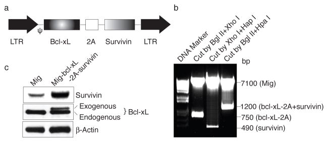
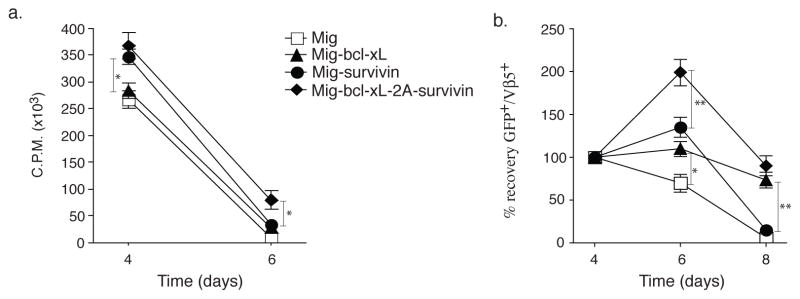
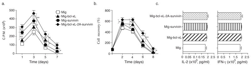
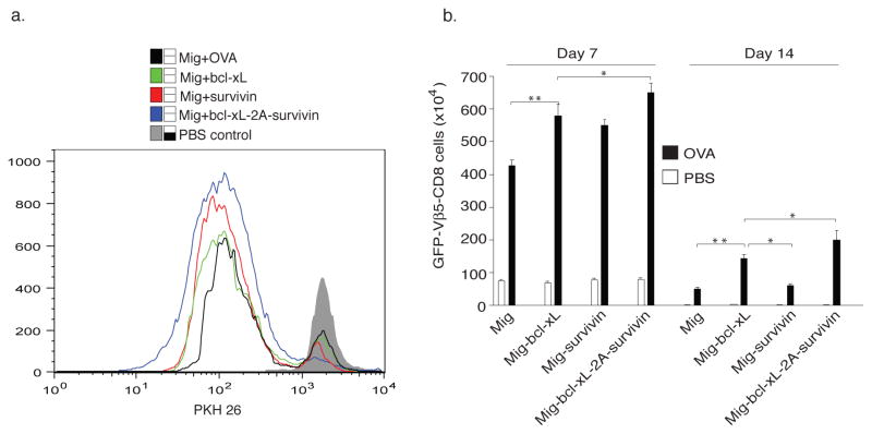
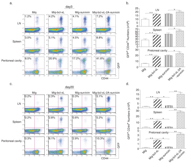
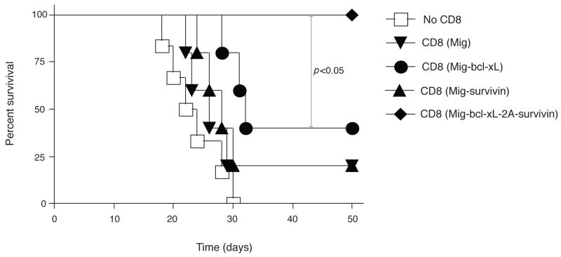
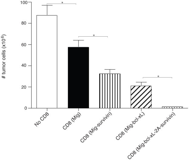
Costimulation regulates multiple cellular processes of T cells inducing proliferation, expansion, and survival. The molecular targets of costimulation might then be useful to augment T cell activities. Two defined targets of costimulatory signals in primary T cells are the anti-apoptotic bcl-2 family molecule Bcl-x(L), and survivin, an inhibitor of apoptosis family member that might regulate both cell division and survival. However, the relative importance of, and relationship between, these molecules in primary T cells is not clear. To understand whether they have overlapping or cooperative functions, we used retrovirus-mediated transduction to introduce Bcl-x(L) and survivin separately, or together linked by a 2A picornavirus self-cleaving peptide, into Ag-responding CD8(+) T cells. We found that CD8(+) effector T cells expressing both Bcl-x(L) and survivin strongly expanded at an early stage and had a long-term survival advantage over cells transduced with either molecule alone. In vivo, with response to tumor-expressed Ag following adoptive T cell transfer, Ag-reactive CD8(+) T cells expressing both Bcl-x(L) and survivin displayed greatly enhanced tumor protective activity compared with CD8(+) T cells expressing either molecule introduced separately. These results indicate that Bcl-x(L) and survivin can critically contribute in a cooperative, nonredundant manner to augment the accumulation and persistence of CD8(+) T cells following encounter with Ag. The data provide new insights into why costimulatory signals might need to be sustained over time and suggest a potential novel approach to augment cellular immunotherapy for cancer.


Figures

Figure 1
Figure 1
Expression of Bcl-xL and Survivin using a 2A gene sequence in primary CD8+ T cells. (a) Schematic representation of the retrovirus construct expressing Bcl-xL and Survivin. Ψ, packaging signal. 2A, picornavirus self-cleaving 2A sequence. (b) Genes of Bcl-xL and Survivin were linked with T2A sequence and were subcloned into Mig vector (Mig-bcl-xL-2A-survivin), which was confirmed by digestion with restriction enzymes, showing fragments of Survivin (~500 bp), Bcl-xL-2A (~750 bp), and bcl-xL-2A-survivin (~1,200 bp). (c) Naive CD8+ T cells from OT-I TCR transgenic mice were stimulated with peptide/APCs. On day 2/3, T cells were transduced with retroviral vectors expressing GFP (Mig), GFP with Bcl-xL (Mig-bcl-xL), GFP with Survivin (Mig-survivin), or GFP with Bcl-xL and Survivin (Mig-bcl-xL-2A-survivin). On day 5 of primary culture, GFP+ CD8+ T cells were sorted, and protein expression of Bcl-xL, Survivin, and β-actin was determined by western blotting. Data are representative of three independent experiments.
Figure 2
Figure 2
Retroviral transduction of Bcl-xL and Survivin promotes passive proliferation and survival of CD8+ T cells in vitro. Naive CD8+ T cells from OT-I TCR transgenic mice were stimulated with peptide/APCs, and transduced on days 2/3 with retroviral vectors expressing GFP, GFP with Bcl-xL, GFP with Survivin, or GFP with Bcl-xL and Survivin, and then recultured without any further stimulation. (a) Primary passive proliferation on day 4 and day 6 were measured in unseparated cultures by pulsing with tritiated thymidine for 20 hr. Data are representative with mean of three independent experiments (* P<0.05, Student’s unpaired t-test). (b) GFP+Vβ5+ T cell recovery normalized to take into account differences in initial transduction efficiency between cultures. Numbers of GFP+ cells present on day 4 were assigned a value of 100%, and numbers surviving on day 6 and day 8 were used to calculate the percentage recovery relative to day 4. Data represent the mean ± S.D. percentage change from three separate experiments (* P<0.05, ** P<0.01, Student’s unpaired t-test). GFP+ CD8+ T cells on day 6 were sorted, and protein expression of Survivin and β-actin was also determined by western blotting, Data are representative of three independent experiments. (c) CD25 expression on day 4 was analyzed by flow cytometry, after gating on live CD8+ GFP+ T cells. Data are representative of three independent experiments. (d) Apoptosis of GFP+ CD8+ T cells on day 6 based on staining of Annexin V and 7-AAD and analyzed by flow cytometry. Data are representative of three independent experiments.
Figure 3
Figure 3
Retroviral transduction of Bcl-xL and Survivin augments the proliferation and survival of CD8+ T cells in secondary responses in vitro. Naive CD8+ T cells from OT-I TCR transgenic mice were stimulated with peptide/APCs. On day 2/3, T cells were transduced with retroviral vectors expressing GFP, GFP with Bcl-xL, GFP with Survivin, or GFP with Bcl-xL and Survivin. On day 5 of primary culture, GFP+ CD8+ T cells were sorted, and restimulated with APCs/peptide. (a) Recall proliferation on day 1 to day 7 measured by pulsing with tritiated thymidine for the last 20 hr. Data are mean cpm ± S.D. from triplicate cultures and are representative of three experiments (* P<0.05, ** P<0.01, Student’s unpaired t-test). (b) Recall survival, based on recovery of GFP+Vβ5+ T cells over time. Cell numbers present on day 0 were assigned a value of 100%, and cell numbers surviving on day 2 to day 8 were used to calculate the percentage recovery. Data represent the mean ± S.D. percentage change from three separate experiments (* P<0.05, Student’s unpaired t-test). GFP+ CD8+ T cells on day 3 were sorted, and protein expression of Survivin and β-actin was also determined by western blotting, Data are representative of three independent experiments. (c) Recall IL-2 and IFN-γ production were measured by ELISA at 40 h. Data are representative of three independent experiments. (d) Apoptosis of GFP+ CD8+ T cells on day 4 based on staining of Annexin V and 7-AAD and analyzed by flow cytometry. Data are representative of three independent experiments.
Figure 4
Figure 4
Retroviral transduction of Bcl-xL and Survivin sustains CD8+ T cell proliferation and long-term survival. Naive CD8+ T cells from OT-I TCR transgenic mice were stimulated with peptide/APCs. On day 2/3, T cells were transduced with retroviral vectors expressing GFP, GFP with Bcl-xL, GFP with Survivin, or GFP with Bcl-xL and Survivin. On day 5 of primary culture, GFP+ CD8+ T cells were sorted, labeled with the dye PKH26, and adoptively transferred into naive recipient mice that were subsequently challenged i.p. with whole OVA protein (100 μg) in PBS (filled bars) or with PBS alone (open bars). (a) Cell division of GFP+ CD8+ T cells on day 3 based on dilution of PKH26. The mean fluorescence intensity of PKH 26 expression, the percentage of diluted PKH 26 in each group and the histogram overlay were shown. Data are representative of three independent experiments. (b) On days 7 and 14, GFP+Vβ5+CD8+ T cells were enumerated from pooled lymph nodes and spleen. Data are mean number of GFP+Vβ5+CD8+ ± S.D. from six individual mice and representative of three independent experiments (* P<0.05, ** P<0.01, Student’s unpaired t-test).
Figure 5
Figure 5
Retroviral transduction of Bcl-xL and Survivin promotes initial CD8+ T cell expansion in vivo in response to tumor antigen. Naive CD8+ T cells from OT-I TCR transgenic mice were stimulated with APCs/peptide. On day 2/3, T cells were transduced with retroviral vectors expressing GFP, GFP with Bcl-xL, GFP with Survivin, or GFP with Bcl-xL and Survivin. On day 5 of primary culture, GFP+ CD8 T cells were sorted and adoptively transferred into naive recipient mice that were subsequently challenged i.p. with EG.7 tumor cells expressing OVA. At different time points, percentage of GFP+CD44+ T cells was analyzed by flow cytometry, after gating on live CD8+ T cells in the lymph nodes, spleen, and peritoneal cavity (a, d). Only tumor cell challenged mice shown. Results are representative of three experiments. Actual numbers of GFP+CD44+ T cells in pooled lymph nodes (LN: inguinal, mesenteric, and paraaortic; top), spleen (middle), and peritoneal cavity (bottom) (b, e). Data are mean number of GFP+CD44+ T cells ± S.D. from six individual mice (* P<0.05, ** P<0.01, Student’s unpaired t-test). At different time points, single-cell suspensions from pooled lymph nodes, spleen, and peritoneal cavity were stimulated with OVA peptide for 7 hours, INF-γ and granzyme B were analyzed by intracellular staining, after gating on live CD8+ GFP+ T cells (c, f). Data are representative of three independent experiments. (a, b, c) Day 3. (d, e, f) Day 20.
Figure 6
Figure 6
Adoptive transfer of Bcl-xL and Survivin transduced CD8+ T cells into tumor-bearing mice augments mouse survival. Naïve CD8+ T cells from OT-I TCR transgenic mice were stimulated with APCs/peptide. On day 2/3, T cells were transduced with retroviral vectors expressing GFP, GFP with Bcl-xL, GFP with Survivin, or GFP with Bcl-xL and Survivin. On day 5 of primary culture, GFP+ CD8+ T cells were sorted and adoptively transferred into naive recipient mice that were subsequently challenged i.p. with EG.7 (a) or EL4 (b) tumor cells. Percent survival was studied for 50 days. Kaplan Meier survival analysis indicated significantly increased survival in mice (n=6) receiving Bcl-xL and Survivin transduced CD8+ T cells compared to control groups (log rank test, P<0.05).
Figure 7
Figure 7
Adoptive cell transfer of Bcl-xL and Survivin transduced CD8+ T cells prevents tumor growth. Naïve CD8+ T cells from OT-I TCR transgenic mice were stimulated with APCs/peptide. On day 2/3, T cells were transduced with retroviral vectors expressing GFP, GFP with Bcl-xL, GFP with Survivin, or GFP with Bcl-xL and Survivin. On day 5 of primary culture, GFP+ CD8+ T cells were sorted and adoptively transferred into naive recipient mice that were subsequently challenged i.p. with EG.7 tumor cells. On day 20, the peritoneal cavity was flushed with 10 ml of ice-cold PBS twice to extract tumor cells and recruited lymphocytes. The total number of cells obtained from each site and the numberof tumor cells from the peritoneal cavity were counted by trypanblue exclusion. Data are mean number of tumor cells ± S.D. from six individual mice (* P<0.05, One-way ANOVA test).

Similar articles

Cited by

References

    1. Croft M. Co-stimulatory members of the TNFR family: keys to effective T-cell immunity? Nat Rev Immunol. 2003;3:609–620. - PubMed
    1. Watts TH. TNF/TNFR family members in costimulation of T cell responses. Annu Rev Immunol. 2005;23:23–68. - PubMed
    1. Yang Y, Liu XK, Nguyen T, Bishop C, Graf D, Dong C. Characterization of B7S3 as a novel negative regulator of T cells. J Immunol. 2007;178:3661–3667. - PubMed
    1. Zang X, Allison JP. To be or not to be B7. J Clin Invest. 2006;116:2590–2593. - PMC - PubMed
    1. Khoury SJ, Sayegh MH. The roles of the new negative T cell costimulatory pathways in regulating autoimmunity. Immunity. 2004;20:529–538. - PubMed

Publication types