. 2009 Jun 16;100(12):1957-65.
doi: 10.1038/sj.bjc.6605060. Epub 2009 May 19.

Inhibition of Notch pathway prevents osteosarcoma growth by cell cycle regulation

Affiliations

Inhibition of Notch pathway prevents osteosarcoma growth by cell cycle regulation

M Tanaka et al. Br J Cancer. .

Abstract

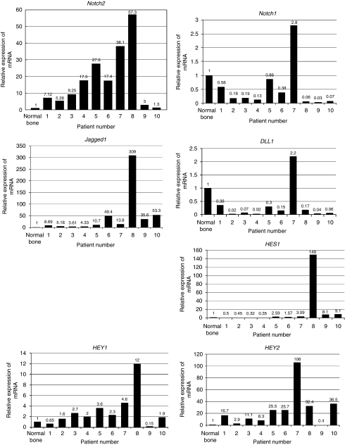
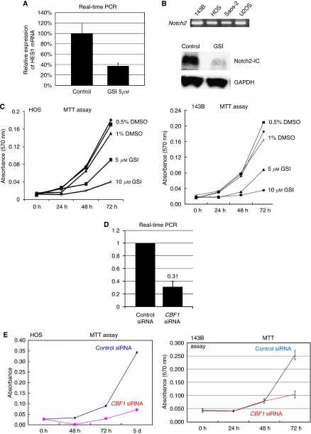
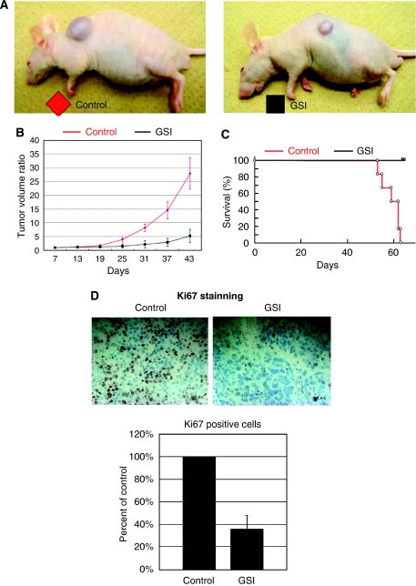
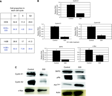
The study shows constitutive activation of the Notch pathway in various types of malignancies. However, it remains unclear how the Notch pathway is involved in the pathogenesis of osteosarcoma. We investigated the expression of the Notch pathway molecules in osteosarcoma biopsy specimens and examined the effect of Notch pathway inhibition. Real-time PCR revealed overexpression of Notch2, Jagged1, HEY1, and HEY2. On the other hand, Notch1 and DLL1 were downregulated in biopsy specimens. Notch pathway inhibition using gamma-secretase inhibitor and CBF1 siRNA slowed the growth of osteosarcomas in vitro. In addition, gamma-secretase inhibitor-treated xenograft models exhibited significantly slower osteosarcoma growth. Cell cycle analysis revealed that gamma-secretase inhibitor promoted G1 arrest. Real-time PCR and western blot revealed that gamma-secretase inhibitor reduced the expression of accelerators of the cell cycle, including cyclin D1, cyclin E1, E2, and SKP2. On the other hand, p21(cip1) protein, a cell cycle suppressor, was upregulated by gamma-secretase inhibitor treatment. These findings suggest that inhibition of Notch pathway suppresses osteosarcoma growth by regulation of cell cycle regulator expression and that the inactivation of the Notch pathway may be a useful approach to the treatment of patients with osteosarcoma.


Figures

Figure 1
Figure 1
Notch pathway molecules are overexpressed in osteosarcoma patient specimens. Total RNA extracted from osteosarcoma biopsy specimens was used for real-time PCR. Ten of ten human biopsy specimens of osteosarcoma increased Notch2 1.3–57.3-fold. Notch1 was decreased 0.03–0.86-fold in 9 of 10 biopsy specimens. Jagged1 was upregulated 3.6–309-fold in 10 of 10 biopsy specimens. In 9 of 10 human biopsy specimens, DLL1 was decreased 0.02–0.35-fold. HES1 was upregulated in 6 of 10 and downregulated in 4 of 10 biopsy specimens. HEY1 was upregulated 1.6–12-fold in 8 of 10 biopsy specimens. HEY2 was upregulated 2.9–106-fold in 9 of 10 biopsy specimens. The comparative Ct (ΔΔCt) method was used to determine fold change in expression using βII-microglobulin. Each sample was run minimally at three concentrations in triplicate.
Figure 2
Figure 2
Inhibition of Notch pathway prevents proliferation of osteosarcoma in vitro. Real-time PCR revealed four human osteosarcoma cell lines that express Notch2 (B). We carried out RT–PCR to determine which concentration of γ-Secretase inhibitor (GSI) X effectively inhibited Notch activity in osteosarcoma cells, and then measured the expression of the Notch pathway target HES1. γ-Secretase inhibitor X at 5 μM reduced mRNA levels of HES1 in 143B cells more than 60% (error bar means s.d.) (A). Western blot analysis showed that 5 μM GSI treatment reduced Notch2 intercellular domain (B). Growth of viable HOS and 143B cells over 3 days was slowed in dose-dependent fashion by GSI X (C). Real-time PCR revealed that siRNA knocked down CBF1 mRNA about 69% (D). Growth of HOS and 143B cells was slowed by CBF1 siRNA (E).
Figure 3
Figure 3
Notch pathway inhibition blocks osteosarcoma xenograft growth in vivo and prolongs survival. In all, 143B cells (1 × 10⁶) were inoculated subcutaneously. Established 143B tumours were measured and then injected with γ-secretase inhibitor (GSI) or DMSO intraperitoneally. The tumour volume at day 7 was set at 1, and tumour volumes at subsequent time points were calculated. γ-Secretase inhibitor significantly inhibited tumour growth at day 31 compared with DMSO. The following decreases in tumour volume were observed in GSI compared with DMSO treatment: day 25: 37.9%; day 31: 26.3%; day 37: 19.7%; and day 43: 18.6% (A and B). Kaplan–Meier survival curves from GSI treatment groups (black) and DMSO control (red). Kaplan–Meier analysis showed that GSI administration conferred a significant survival benefit (C; n=6, P<0.05). Immunohistochemical examination of ki67 was carried out in xenograft tumours. Ki67 staining revealed that proliferation of osteosarcoma cells was decreased by GSI treatment. The number of Ki67-positive cells was decreased to 36% of control revel by GSI administration at day 35 (D; error bar means s.d.).
Figure 4
Figure 4
Notch pathway inhibition promotes G1 arrest. HOS and143B cells were treated with 5 μM GSI. After 48-h treatment, cells were collected and subjected to cell cycle analysis. When HOS cells were cultured without GSI, 54.6% of cells were in G1 phase. On the other hand, when cultured with GSI, 64.8% of cells were in G1 phase. In the case of 143B cells cultured without GSI, 39.8% of cells were in G1 phase, whereas 53.3% of cells were in G1 phase when treated with GSI (A). Real-time PCR was carried out to quantify mRNAs of cell cycle-related genes. A 24-h treatment with GSI reduced the levels of cyclin D, cyclin E1, E2, SKP2, and c-Myc transcription (error bar means s.d.; B). Western blot analysis of the levels of cell cycle-related genes. 48 h treatment with GSI reduced the levels of expression of cyclin E1, cyclin E2, c-Myc, pRb, and SKP2 proteins. Expression of P21cip1 protein was upregulated by GSI treatment. The experiment was triplicate with similar results (C; GSI: 5 μM).

References

    1. Artavanis-Tsakonas S, Rand MD, Lake RJ (1999) Notch signaling: cell fate control and signal integration in development. Science 284: 770–776 - PubMed
    1. Bettenhausen B, Hrabe de Angelis M, Simon D, Guenet JL, Gossler A (1995) Transient and restricted expression during mouse embryogenesis of Dll1, a murine gene closely related to Drosophila delta. Development 121: 2407–2418 - PubMed
    1. Bin Hafeez B, Adhami VM, Asim M, Siddiqui IA, Bhat KM, Zhong W, Saleem M, Din M, Setaluri V, Mukhtar H (2009) Targeted knockdown of Notch1 inhibits invasion of human prostate cancer cells concomitant with inhibition of matrix metalloproteinase-9 and urokinase plasminogen activator. Clin Cancer Res 15: 452–459 - PMC - PubMed
    1. Curry CL, Reed LL, Golde TE, Miele L, Nickoloff BJ, Foreman KE (2005) Gamma secretase inhibitor blocks Notch activation and induces apoptosis in Kaposi's sarcoma tumor cells. Oncogene 24: 6333–6344 - PubMed
    1. Dang TP, Gazdar AF, Virmani AK, Sepetavec T, Hande KR, Minna JD, Roberts JR, Carbone DP (2000) Chromosome 19 translocation, overexpression of Notch3, and human lung cancer. J Natl Cancer Inst 92: 1355–1357 - PubMed

Publication types

MeSH terms

Substances