Case Reports
. 2009 Nov;17(11):1411-6.
doi: 10.1038/ejhg.2009.76. Epub 2009 May 20.

Homozygosity for a null allele of COL3A1 results in recessive Ehlers-Danlos syndrome

Affiliations
Case Reports

Homozygosity for a null allele of COL3A1 results in recessive Ehlers-Danlos syndrome

Aurélie Plancke et al. Eur J Hum Genet. 2009 Nov.

Abstract

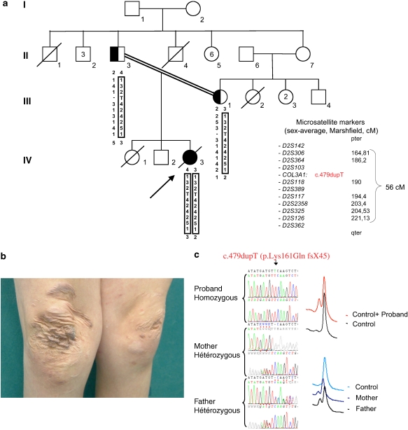
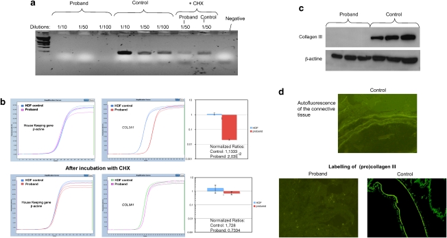
So far, mutations in the human COL3A1 gene have been associated with the predominantly inherited Ehlers-Danlos syndrome (EDS), vascular type. Genotype-phenotype correlation perspectives collapsed, as haploinsufficiency, which was long suggested to confer a milder or unrecognized phenotype, was reported in four patients with a phenotype similar to that of vascular EDS. Here, we study a case of recessive EDS in a young consanguineous girl of healthy parents. She fulfilled the vascular EDS criteria for laboratory testing. Total sequencing of COL3A1 cDNA identified a homozygous nucleotide duplication (c.479dupT) resulting in a premature termination codon (p.Lys161GlnfsX45). Studies in genomic DNA showed that this mutation was inherited from each parent. The expression analysis (RT-PCR, quantitative-PCR, immunohistochemistry, WB) showed strong mRNA decay and an absence of type III collagen in the proband. The expected COL3A1 haploinsufficiency in her healthy ascendants did not lead to the manifestations of vascular EDS. This case provides evidence of a stochastic effect of COL3A1 haploinsufficiency in humans, which could be explained by the relation between nonsense-mediated mRNA decay efficiency and the resulting dominant-negative effect depending on the position of the mutation and/or modifying factors. It opens up new perspectives for the understanding of COL3A1 genotype-phenotype correlations, which is required while considering targeted therapy.


Figures

Figure 1
Figure 1
Pedigree, haplotype, clinical assessment and mutation status. (a) Haplotype study showing a large 56 cM chromosome 2 region (black boxed), inherited by descent in the proband (IV.3). Markers were purchased from Applied Biosystems, Courtaboeuf, France. Genotyping and analysis were carried out according to the manufacturer's instructions. Presence of the mutation, c.479dupT, is represented as a T in haplotype. (b) Characteristic Ehlers–Danlos syndrome (EDS) skin lesions of the Proband. (c) Electropherograms of COL3A1 exon 5 genomic sequences around the c.479dupT, and its detection with denaturing high-performance liquid chromatography (dHPLC, Transgenomic, Courtaboeuf, France) in genomic DNA (additional details in online-only material).
Figure 2
Figure 2
Expression analysis in proband. (a) RT-PCR products separated on an agarose gel. Amplification of 25 cycles of COL3A1 exons 1–2 in the cDNA of a control individual (human dermal fibroblasts (HDF)) and the proband at different dilutions (1/10; 1/50 and 1/100) and with or without cycloheximide (CHX) treatment (only 1/50 dilution is shown). Although a specific transcript is detected in the control, none is detected in the proband without CHX treatment. (b) Real-time RT-PCR. PCR products of COL3A1 exons 5–8 amplicon (primers in online only material) were quantified using the LightCycler 480 system with SYBR Green I Master Mix according to the manufacturer's instructions (Roche, Mannheim, Germany). The expression level of COL3A1 was normalized to that of housekeeping genes, β-actin and GAPDH (Eurogentec, Angers, France). A calibrator sample (HDF) was used and experiments were repeated thrice. In each experiment, the samples were run in triplicates. The COL3A1 expression ratio was calculated using the E-Method (Roche, Eurogentec, Angers, France). (c) Western blot analysis: Immunodetection of Type III procollagen by an antibody against the N-telopeptide (S17, Santa Cruz, Santa Cruz, CA, USA) and by an antibody against the reference gene, β-actin (AC-15, Santa Cruz). We loaded 25, 40 and 60 μg of total proteins that were extracted from both the medium and the cell layer of the skin-cultured fibroblast from the proband and from the control. No (pro)collagen III was detected in the proband. (d) Immunohistochemistry: in situ detection of (pro)collagen III in frozen jejunum samples of the proband and control (48 years old, Caucasian female). In the proband, only autofluorescence of the connective tissue is detected after labeling with the S17 antibody (Santa Cruz), whereas the control showed intense labeling (concordant data were obtained with antibodies against the full-length collagen III triple-helical domain (FH-7A, Santa Cruz; data not shown)).

References

    1. Beighton P, De Paepe A, Steinmann B, Tsipouras P, Wenstrup RJ. Ehlers-Danlos syndromes: revised nosology, Villefranche, 1997. Am J Med Genet. 1998;77:31–37. - PubMed
    1. Pepin M, Schwarze U, Superti-Furga A, Byers PH. Clinical and genetic features of Ehlers-Danlos syndrome type IV, the vascular type. N Engl J Med. 2000;342:673–680. - PubMed
    1. Prockop DJ, Kivirikko KI. Collagens: molecular biology, diseases, and potentials for therapy. Annu Rev Biochem. 1995;64:403–434. - PubMed
    1. Myllyharju J, Kivirikko KI. Collagens, modifying enzymes and their mutations in humans, flies and worms. Trends Genet. 2004;20:33–43. - PubMed
    1. Dalgleish R. The human collagen mutation database 1998. Nucleic Acids Res. 1998;26:253–255. - PMC - PubMed

Publication types

Substances