. 2009 Nov 1;77(2):477-89.
doi: 10.1002/prot.22463.

Targeted molecular dynamics reveals overall common conformational changes upon hybrid domain swing-out in beta3 integrins

Affiliations

Targeted molecular dynamics reveals overall common conformational changes upon hybrid domain swing-out in beta3 integrins

Davide Provasi et al. Proteins. .

Abstract

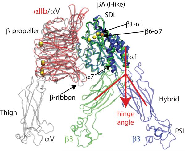
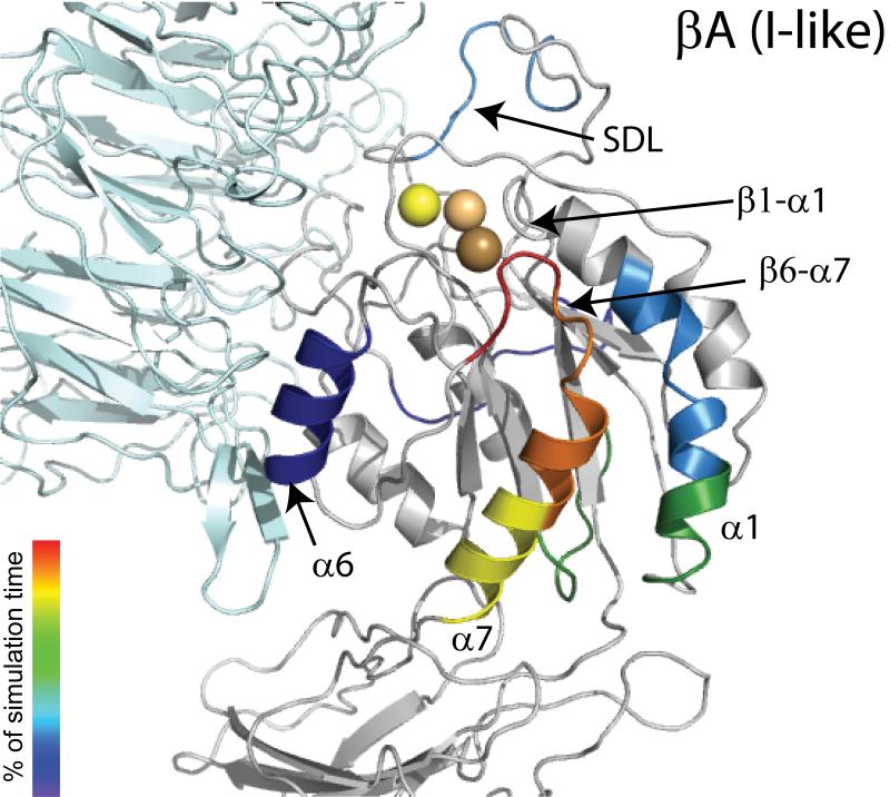
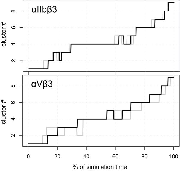
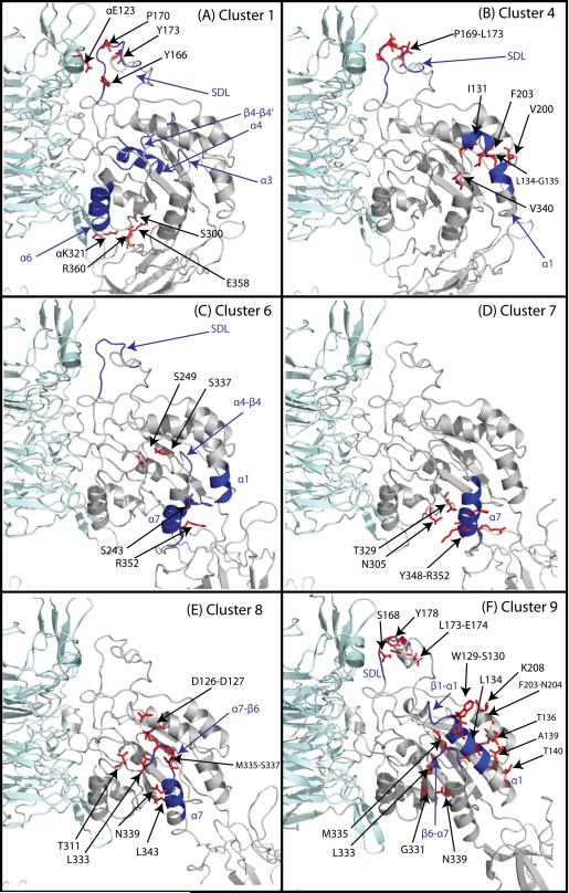
The beta3 integrin family members alphaIIbeta3 and alphaVbeta3 signal bidirectionally through long-range allosteric changes, including a transition from a bent unliganded-closed low-affinity state to an extended liganded-open high-affinity state. To obtain an atomic-level description of this transition in an explicit solvent, we carried out targeted molecular dynamics simulations of the headpieces of alphaIIbeta3 and alphaVbeta3 integrins. Although minor differences were observed between these receptors, our results suggest a common transition pathway in which the hybrid domain swing-out is accompanied by conformational changes within the beta3 betaA (I-like) domain that propagate through the alpha7 helix C-terminus, and are followed by the alpha7 helix downward motion and the opening of the beta6-alpha7 loop. Breaking of contact interactions between the beta6-alpha7 loop and the alpha1 helix N-terminus results in helix straightening, internal rearrangements of the specificity determining loop (SDL), movement of the beta1-alpha1 loop toward the metal ion dependent adhesion site (MIDAS), and final changes at the interfaces between the beta3 betaA (I-like) domain and either the hybrid or the alpha beta-propeller domains. Taken together, our results suggest novel testable hypotheses of intradomain and interdomain interactions responsible for beta3 integrin activation.


Figures

Figure 1
Figure 1. System Setup Used in the TMD Simulations of the Unliganded-closed to the Liganded-open States of β3 Integrins
Comparison between the crystal structures of integrin αIIbβ3 in its liganded-open headpiece conformation (PDBID: 1TY6; color red for the αIIb β-propeller, blue for β3, and yellow for metal ions) and integrin αVβ3 in its unliganded-closed state (PDBID: 1U8C; color gray for both the β-propeller and thigh domains of αV, green for β3, and orange for metal ions). The regions of the β3 βA (I-like) domain that show the largest conformational differences are labeled and indicated with arrows. The relative opening of the hinge angle between the β3 βA (I-like) and hybrid domains that describes the swing-out motion of the hybrid domain is indicated by red lines and the red arrow.
Figure 2
Figure 2. RMSD to the Target Configuration Calculated During the RP-TMD Runs
The distance from the TMD target is illustrated for both αIIbβ3 (upper panel) and the αVβ3 (lower panel) integrins. The average over the two statistically independent simulations we carried out for each integrin system is represented as a solid line, in black for the forward (unliganded-closed to liganded-open) simulation and in gray for the backward (liganded-open to unliganded-closed) one, along with 95% confidence interval. The insets of the two panels illustrate the behavior of the hinge angle (see text) with the same color coding used above.
Figure 3
Figure 3. Sequence of Events During the RP-TMD Simulations of the Transition from Unliganded-Closed to the Liganded-Open States of αIIbβ3 integrin
Structure of the unbound βA (I-like) domain showing the different secondary structure elements colored according to the order of significant conformational changes occurring during simulations with respect to the starting conformation. The α subunit is represented in cyan while SyMBS, MIDAS, and ADMIDAS are shown in light yellow, light orange, and light brown CPKs, respectively. Color coding for conformational changes at the very end of the simulation (after 90% simulation time), including the movement of β1-α1 towards the MIDAS, α1 helix straightening, internal rearrangements of SDL, and final changes at the interfaces between the β3 βA (I-like) domain and either the hybrid or the α β-propeller domains, is omitted for clarity.
Figure 4
Figure 4. Dynamics of β3 Integrins in Cluster Space
Conformational evolution of the integrin systems among the clusters defined by the contact matrix analysis of the RP-TMD simulations as a function of the elapsed fraction of simulation time. The upper and lower panels show the sequence of secondary structure changes in the β3 βA (I-like) domain of integrins αIIbβ3 and αVβ3, respectively, as defined by conformational clustering of their forward (unliganded-closed to liganded-open states; black line) and backward (liganded-open to unliganded-closed states; gray line) trajectories. To make the comparison easier, the curves corresponding to the backward simulations (gray color) have been reversed, so that the abscissa represents the fraction of time from the end of the simulation.
Figure 5
Figure 5. Representative Conformations of the Most Relevant Clusters of Integrin αIIbβ3
The most relevant clusters obtained by RP-TMD are defined as the clusters whose conformations show significant differences (RMSD > 4Å). The αIIb subunit is depicted in cyan, and the β3 subunit in gray. Some of the relevant residues involved in contact breaking and formation in the transitions from one cluster to the other are depicted as red sticks. The secondary structure motifs that show significant conformational differences among sequential clusters are depicted in blue. The numbers refer to the residue index in the β3 subunit, unless otherwise indicated.

References

    1. Shattil SJ, Newman PJ. Integrins: dynamic scaffolds for adhesion and signaling in platelets. Blood. 2004;104(6):1606–1615. - PubMed
    1. Byzova TV, Rabbani R, D’Souza SE, Plow EF. Role of integrin alpha(v)beta3 in vascular biology. Thromb Haemost. 1998;80(5):726–734. - PubMed
    1. Fitzgerald LA, Poncz M, Steiner B, Rall SC, Jr., Bennett JS, Phillips DR. Comparison of cDNA-derived protein sequences of the human fibronectin and vitronectin receptor alpha-subunits and platelet glycoprotein IIb. Biochemistry. 1987;26(25):8158–8165. - PubMed
    1. Blue R, Murcia M, Karan C, Jirouskova M, Coller BS. Application of high-throughput screening to identify a novel alphaIIb-specific small- molecule inhibitor of alphaIIbbeta3-mediated platelet interaction with fibrinogen. Blood. 2008;111(3):1248–1256. - PMC - PubMed
    1. Smith JW, Ruggeri ZM, Kunicki TJ, Cheresh DA. Interaction of integrins alpha v beta 3 and glycoprotein IIb-IIIa with fibrinogen. Differential peptide recognition accounts for distinct binding sites. J Biol Chem. 1990;265(21):12267–12271. - PubMed

Publication types

LinkOut - more resources