. 2009 Jul 7;48(26):6191-201.
doi: 10.1021/bi900629e.

Structural basis for the sugar nucleotide and acyl-chain selectivity of Leptospira interrogans LpxA

Affiliations

Structural basis for the sugar nucleotide and acyl-chain selectivity of Leptospira interrogans LpxA

Lori I Robins et al. Biochemistry. .

Abstract

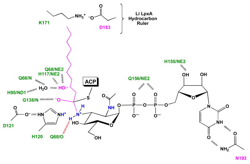
The first step of lipid A biosynthesis is catalyzed by LpxA in Escherichia coli (EcLpxA), an acyltransferase selective for UDP-GlcNAc and R-3-hydroxymyristoyl-acyl carrier protein (ACP). Leptospira interrogans LpxA (LiLpxA) is extremely selective for R-3-hydroxylauroyl-ACP and an analogue of UDP-GlcNAc, designated UDP-GlcNAc3N, in which NH(2) replaces the GlcNAc 3-OH group. EcLpxA does not discriminate between UDP-GlcNAc and UDP-GlcNAc3N; however, E. coli does not make UDP-GlcNAc3N. With LiLpxA, R-3-hydroxylauroyl-methylphosphopantetheine efficiently substitutes for R-3-hydroxylauroyl-ACP. We now present crystal structures of free LiLpxA and its complexes with its product UDP-3-N-(R-3-hydroxylauroyl)-GlcNAc3N and with its substrate R-3-hydroxylauroyl-methylphosphopantetheine. The positions of the acyl chains of the R-3-hydroxylauroyl-methylphosphopantetheine and the UDP-3-N-(R-3-hydroxylauroyl)-GlcNAc3N are almost identical and are similar to that of the acyl chain in the EcLpxA/UDP-3-O-(R-3-hydroxymyristoyl)-GlcNAc complex. The selectivity of LiLpxA for UDP-GlcNAc3N may be explained by the orientation of the backbone carbonyl group of Q68, which differs by approximately 82 degrees from the corresponding Q73 carbonyl group in EcLpxA. This arrangement provides an extra hydrogen-bond acceptor for the 3-NH(2) group of UDP-GlcNAc3N in LiLpxA. The R-3-hydroxylauroyl selectivity of LiLpxA is explained by the position of the K171 side chain, which limits the length of the acyl-chain-binding groove. Our results support the role of LiLpxA H120 (which corresponds to EcLpxA H125) as the catalytic base and provide the first structural information about the orientation of the phosphopantetheine moiety during LpxA catalysis.


Figures

Figure 1
Figure 1. Substrate selectivity of LiLpxA versus EcLpxA and the origin of UDP-GlcNAc3N
Panel A. Biosynthesis of UDP-GlcNAc3N, substrate selectivity of LiLpxA and structure of L. interrogans lipid A (6-8). The R-3-hydroxylauroyl (R-3-OHC12) chain is shown in magenta both in the LiLpxA product and in the lipid A generated by L. interrogans. Panel B. Selectivity of EcLpxA and structure of E. coli lipid A (12). The R-3-hydroxymyristate (R-3-OHC14) chain is shown in green. The E. coli chromosome does not encode GnnA and GnnB, but EcLpxA can utilize UDP-GlcNAc3N as efficiently as UDP-GlcNAc in vitro (7).
Figure 2
Figure 2. Asymmetric units and backbone structures of free LiLpxA versus EcLpxA
Panel A. There are three chains (light blue cartoon diagrams) in the LiLpxA asymmetric unit, each containing 259 residues. The N-terminal domain of each chain is folded into a left-handed parallel β-helix. The C-terminal domain consists of four α-helices. Black arrows in chain A indicate origins of loops inserted within the β-helix domain. Panel B. Bottom up and side views of the EcLpxA asymmetric unit (14), which consists of a single chain of 262 amino acids (red cartoon diagram). Loops of similar position and size (black arrows) to those seen in LiLpxA are present in the β-helix domain of EcLpxA. The second loop of LiLpxA is smaller and oriented differently than that of EcLpxA. Panel C. Superposition of EcLpxA (14) and chain A of LiLpxA.
Figure 3
Figure 3. Structure of the biological LiLpxA homotrimer and position of active site residues in the absence of ligands
Panel A. Side view of the free LiLpxA biological trimer (pale green, slate blue and light pink cartoons), generated from chain A of the asymmetric unit. The EcLpxA monomer of its asymmetric unit (brick red) (14) is superimposed on one LiLpxA subunit for comparison of the active site region. Panel B. Close up view of superimposed active site residues. LiLpxA H120 (corresponding to EcLpxA H125) is the catalytic base (Schemes 1 and 2). Q156 side chain of LiLpxA is displaced from that of the corresponding EcLpxA residue Q161. The backbone O atom of Q68 in LiLpxA is rotated relative to its counterpart in Q73 of EcLpxA.
Figure 4
Figure 4. R-3-hydroxylauroyl-methylphosphopantetheine as an acyl donor substrate for LiLpxA
In these assays, the concentration of UDP-GlcNAc3N was 10 μM. Panel A. Near equivalence of 10 μM R-3-hydroxylauroyl-ACP and 50 μM R-3-hydroxylauroyl-methylphosphopantetheine as donors. Panel B. Lack of activity of R-3-hydroxylauroyl-coenzyme A. Abbreviations: CoA, coenzyme A; MePPan, methyl-phosphopantetheine; R-3-OHC12-, R-3-hydroxylauroyl-.
Figure 5
Figure 5. Asymmetric unit and ligand electron densities in the LiLpxA/UDP-3-N-(R-3-hydroxylauroyl)-GlcNAc3N complex
The UDP-3-N-(R-3-hydroxylauroyl)-GlcNAc3N ligands (space filling with yellow carbons) are shown together with their corresponding LiLpxA chains in the asymmetric unit (light blue cartoons). The actual electron density of each ligand (light grey mesh) is superimposed on an expanded stick model of UDP-3-N-(R-3-hydroxylauroyl)-GlcNAc3N in the best calculated conformation (carbons in yellow) next to its corresponding LiLpxA protein chain. The final 2FoFc electron density map is contoured at 1 σ around each ligand.
Figure 6
Figure 6. Position of the Q68 backbone O atom in the LiLpxA/UDP-3-N-(R-3-hydroxylauroyl)-GlcNAc3N complex
Panel A. Ribbon diagram of the biological LiLpxA homotrimer (generated from monomer A of the asymmetric unit) with a space-filling model of the bound product UDP-3-N-(R-3-hydroxylauroyl)-GlcNAc3N. The enzyme subunits of the biological homotrimer are colored in pale green, slate blue and light pink. For UDP-3-N-(R-3-hydroxylauroyl)-GlcNAc3N, the carbon atoms are yellow, the oxygen atoms red, the nitrogen atoms blue, and the phosphorous atoms orange. Panel B. The product complex viewed from the top down. Panel C. Close up of the positions of key active site residues in LiLpxA in relation to bound UDP-3-N-(R-3-hydroxylauroyl)-GlcNAc3N. The color scheme for the ligand and protein is the same as above. Key hydrogen bonds are indicated with black dashes. The NE2 atom of H120 is the proposed catalytic base when catalysis proceeds in the forward direction (Scheme 2), and the backbone N atom of G138 is proposed the oxyanion hole. The ordered water molecule (red) that is hydrogen bonded to the R-3-OH group of the product is seen in all three LiLpxA chains of the asymmetric unit (not shown). Panels D. Active site residues in the LiLpxA/UDP-3-N-(R-3-hydroxylauroyl)-GlcNAc3N complex (chain A) versus the EcLpxA/UDP-3-O-(R-3-hydroxymyristoyl)-GlcNAc complex (12). The color scheme for LiLpxA and its ligand is the same as above. The color scheme for the E. coli protein and its ligand is also the same, except that all carbon atoms are grey. The ordered water molecule in the E. coli complex is cyan.
Figure 7
Figure 7. Asymmetric unit and ligand electron densities in the LiLpxA/R-3-hydroxylauroyl-methylphosphopantetheine complex
Panel A. Covalent structure of synthetic R-3-hydroxylauroyl-methylphosphopantetheine. Panel B. The R-3-hydroxylauroyl-methylphosphopantetheine ligands (space filling models with carbons in cyan) are shown together with their corresponding LiLpxA chains in the asymmetric unit (light blue cartoons). The actual electron density of each ligand (light grey mesh) is superimposed on an expanded stick model of the R-3-hydroxylauroyl-methylphosphopantetheine ligand in the best, calculated conformations (carbons in cyan) next to its corresponding LiLpxA protein chain. The conformations and positions of the hydroxyacyl chains, the thioester linkages, and the β-mercaptoethylamine groups are very similar in each of the three ligands, and their electron densities are strong. The β-alanine, pantoic acids and methyl phosphate groups are modeled in two conformations, and their electron densities are weak. The final 2FoFc electron density map is contoured at 1 σ around each ligand. Thermal motions were analyzed for the donor but not the product complex using TLSMD (41).
Figure 8
Figure 8. Position of the R-3-hydroxylauroyl-methylphosphopantetheine donor substrate at the LiLpxA active site
Panel A. Ribbon diagram of the LiLpxA biological homotrimer with a space-filling model of the bound donor substrate R-3-hydroxylauroyl-methylphosphopantetheine, modeled in two conformations. The subunits of the biological LiLpxA homotrimer (generated from chain A of the asymmetric unit) are colored as in Fig. 6A. The atoms of R-3-hydroxylauroyl-methylphosphopantetheine are colored as follows: carbon cyan, oxygen red, nitrogen blue, sulfur yellow-orange, and phosphorous orange. Panel B. Close-up view of the alternative conformations of the R-3-hydroxylauroyl-methylphosphopantetheine ligand. Abbreviation: Methyl-P, methyl phosphate residue. Panel C. Positions of key active site residues in LiLpxA in relation to bound R-3-hydroxylauroyl-methylphosphopantetheine. The color scheme for the ligand is the same as above. The green and magenta carbons of LiLpxA side chains correspond to the similarly colored ribbons in Panel A to distinguish the subunits of the biological LiLpxA trimer. Key hydrogen bonds are indicated with black dashes. The backbone N atom of G138 is proposed the oxyanion hole. The ordered water molecule (red) that is hydrogen bonded both to the R-3-OH group is seen in all three LiLpxA monomers of the asymmetric unit (not shown) and is the same as in the product complex. The hydrophobic cleft that defines the acyl chain binding site is highlighted by the transparent spheres. Panel D. Superposition of Conformation 1 of the bound donor substrate (cyan carbons) and the bound product (yellow carbons), each associated with chain A of their respective asymmetric units. Abbreviations: Methyl-P, methyl phosphate residue; R-3-OHC12-, R-3-hydroxylauroyl-.
Scheme 1
Scheme 1
Proposed catalytic mechanism of EcLpxA and roles of key residues in substrate binding.
Scheme 2
Scheme 2
Proposed catalytic mechanism of LiLpxA and roles of key residues in substrate binding. The critical H-bond formed between the 3-NH2 group of the acceptor substrate and the backbone O-atom of Q68 is shown in red.

References

    1. Levett PN. Leptospirosis. Clin Microbiol Rev. 2001;14:296–326. - PMC - PubMed
    1. Farr RW. Leptospirosis. Clin Infect Dis. 1995;21:1–6. - PubMed
    1. Ren SX, Fu G, Jiang XG, Zeng R, Miao YG, Xu H, Zhang YX, Xiong H, Lu G, Lu LF, Jiang HQ, Jia J, Tu YF, Jiang JX, Gu WY, Zhang YQ, Cai Z, Sheng HH, Yin HF, Zhang Y, Zhu GF, Wan M, Huang HL, Qian Z, Wang SY, Ma W, Yao ZJ, Shen Y, Qiang BQ, Xia QC, Guo XK, Danchin A, Saint Girons I, Somerville RL, Wen YM, Shi MH, Chen Z, Xu JG, Zhao GP. Unique physiological and pathogenic features of Leptospira interrogans revealed by whole-genome sequencing. Nature. 2003;422:888–893. - PubMed
    1. Bulach DM, Kalambaheti T, de la Pena-Moctezuma A, Adler B. Lipopolysaccharide biosynthesis in Leptospira. J Mol Microbiol Biotechnol. 2000;2:375–380. - PubMed
    1. Weckesser J, Mayer H. Different lipid A types in lipopolysaccharides of phototrophic and related non-phototrophic bacteria. FEMS Microbiol Rev. 1988;4:143–153. - PubMed

Publication types

MeSH terms

Associated data

LinkOut - more resources