. 2009 May 20:4:18; discussion 18.
doi: 10.1186/1745-6150-4-18.

Mapping the sequence mutations of the 2009 H1N1 influenza A virus neuraminidase relative to drug and antibody binding sites

Affiliations

Mapping the sequence mutations of the 2009 H1N1 influenza A virus neuraminidase relative to drug and antibody binding sites

Sebastian Maurer-Stroh et al. Biol Direct. .

Abstract

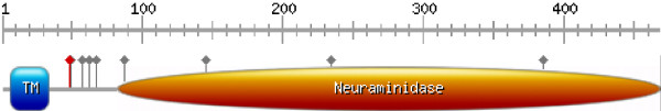
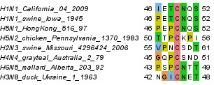
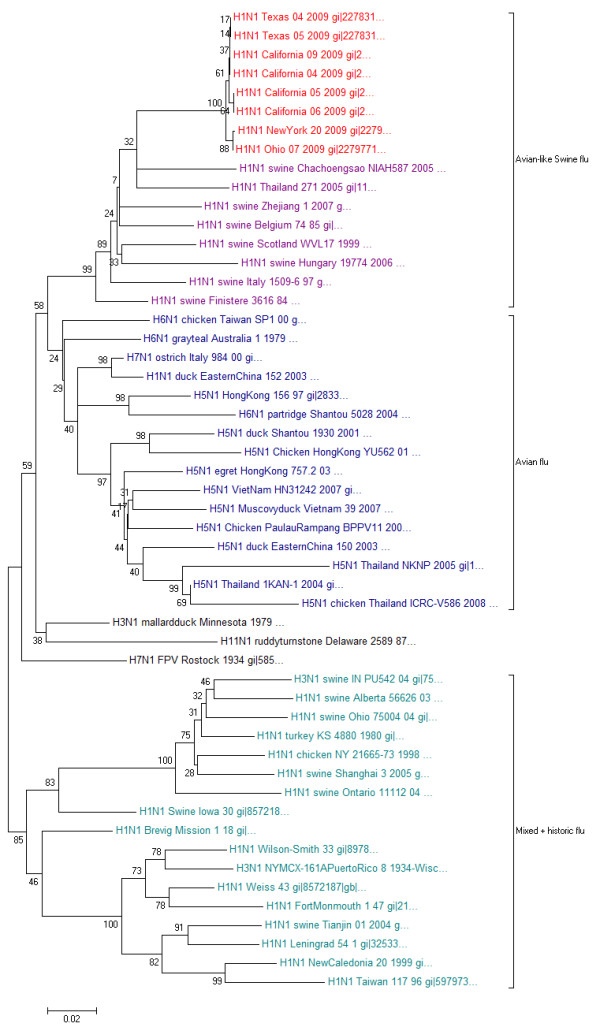
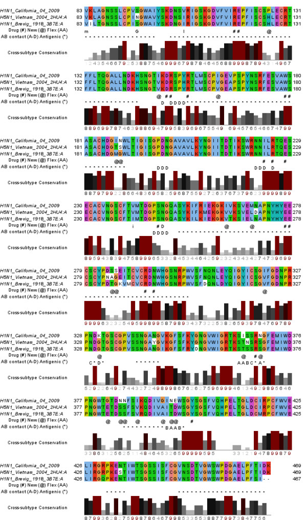
In this work, we study the consequences of sequence variations of the "2009 H1N1" (swine or Mexican flu) influenza A virus strain neuraminidase for drug treatment and vaccination. We find that it is phylogenetically more closely related to European H1N1 swine flu and H5N1 avian flu rather than to the H1N1 counterparts in the Americas. Homology-based 3D structure modeling reveals that the novel mutations are preferentially located at the protein surface and do not interfere with the active site. The latter is the binding cavity for 3 currently used neuraminidase inhibitors: oseltamivir (Tamiflu), zanamivir (Relenza) and peramivir; thus, the drugs should remain effective for treatment. However, the antigenic regions of the neuraminidase relevant for vaccine development, serological typing and passive antibody treatment can differ from those of previous strains and already vary among patients.

Reviewers: This article was reviewed by Sandor Pongor and L. Aravind.


Figures

Figure 1
Figure 1
Domain architecture (drawn with ). Besides the labelled domains (TM ... transmembrane), grey lollipops indicate known and putative glycosylation sites and the red lollipop marks the conserved cysteine shown in Figure 2.
Figure 2
Figure 2
Representative alignment of the sequence environment of the conserved cysteine C49 that could either serve for intermolecular disulfide bridges or as palmitoylation site.
Figure 3
Figure 3
Phylogenetic tree of neuraminidase protein sequences of the N1 subtype family.
Figure 4
Figure 4
A) Surface representation of the structural model of the neuraminidase domain of the new strain in complex with zanamivir. Coloring is based on sequence conservation over all NA subtypes. Grey means no conservation. Other colors are according to physical properties: yellow ... hydrophobic, green ... polar, blue ... positive charge, red ... negative charge. Color intensities are proportional to strength of conservation. B) Mapping of new mutations to structure. Cyan colored residues are mutations at typical antibody recognition sites. Blue residues indicate differences to both the H5N1 avian flu as well as H1N1 from the 1918 Spanish flu. Yellow residues are intra-strain variations occurring in multiple patients of the 2009 H1N1 outbreak and orange if they have only been found in isolated patients, so far. Note that the intra-strain variation N248D is colored cyan since it is part of the antibody recognition site. The backbone of the antibody recognition sites is colored green and the bound drug and 3 calcium ions are shown in red.
Figure 5
Figure 5
Alignment of the NA domain of the 2009 H1N1 strain with the sequences in crystal structures of H5N1 avian flu as well as H1N1 from the 1918 Spanish flu. Residues within 3 Å of the bound drug are indicated with "#", while residues that are different in the new strain compared to both other structures are marked with "@".Intra-strain variation (Flex [AA] in the first annotation line) is displayed as the respective mutated residue in capital letters if found in multiple patients (e.g. D for the N248D substitution) or lower-case (e.g. "i" for V241I) for single occurrences. In the second annotation row, antigenic regions are labelled as "*". Residues with < 3 Å contact to antibodies are labelled "A" for interactions derived from PDB:1ncb, "B" from both PDB:1ncb and PDB:1nmb, "C" from PDB:1nmb and "D" from PDB:2aep.

References

    1. Butler D. Swine flu goes global. Nature. 2009;458:1082–1083. - PubMed
    1. Cohen J, Enserink M. Infectious diseases. As swine flu circles globe, scientists grapple with basic questions. Science. 2009;324:572–573. - PubMed
    1. Vogel G. Public health. SARS and bird flu veteran to take WHO helm. Science. 2006;314:1067. - PubMed
    1. Ooi HS, Kwo CY, Wildpaner M, Sirota FL, Eisenhaber B, Maurer-Stroh S, Wong WC, Schleiffer A, Eisenhaber F, Schneider G. ANNIE: integrated de novo protein sequence annotation. Nucleic Acids Res. 2009 - PMC - PubMed
    1. Eisenhaber F. Prediction of Protein Function: Two Basic Concepts and One Practical Recipe. In: Eisenhaber F, editor. Discovering biomolecular mechanisms with computational biology. Georgetown: Landes Biosciences and Eurekah.com; 2006. pp. 39–54.

MeSH terms

LinkOut - more resources